Co-Immunoprecipitation (Co-IP) Technical
Background
Protein-protein interactions play important roles in both inter- and intra-cellular signaling [1]. Among the various methods of detecting protein-protein interactions, co-IP is cost-saving, easy to handle, and can be applied to various types of sample, such as plant cells [4], mammalian cells [5] yeast [2], and nematode tissue [3].
Experimental outline
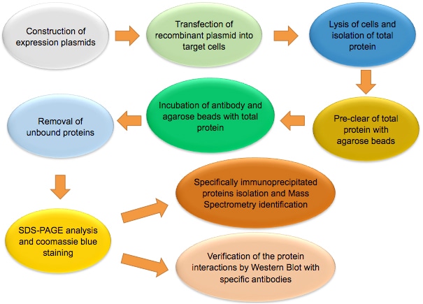
The figure below shows the workflow of co-IP.
Note: Co-IP is a multi-step assay. Experiment should be performed under non-denaturing conditions. Not only high quality reagents, but also careful planning and rigorous operation are needed to obtain good result. Each step of the experiment, from sample preparation, antibody-beads incubation, to co-IP elution, should be tightly controlled to ensure success of the experiment.
Protocol for co-IP to detect protein-protein interaction
1. Recombinant expression plasmid construction
- i. Confirm correct DNA sequence information of the target protein.
- ii. Select proper expression plasmid based on the property of the target protein.
- iii. Insert the target protein in frame with the expression vector to generate recombinant expression plasmid.
Please refer to the relating protocols on our website for the following expression plasmid transformation, colony screening and positive colony verification steps.
2. Transfection into target cells
- i. Amplify and purify the recombinant plasmid after validation of positive clone.
- ii. Transfect the recombinant expression plasmid into target cells.
Cell type used for transfection is determined according to the requirement of the experiment.
3. Cell lysis and total protein isolation
- i. Harvest the cells 24-48 h after transfection.
- ii. Wash the cells with ice-cold phosphate buffer saline (PBS) and centrifuge to collect pellets. Repeat once.
- iii. Gently lyse the cells for 30 min on ice using freshly made proteinase containing lysis buffer.
- iv. Centrifuge the cell lysate at 4 °C and remove cell debris.
Note: In order to maintain the physiological state of the proteins, manipulation of the cell lysis should be under low temperature, the cell lysis buffer should contain mild non-ionic detergent, proteinase inhibitor, and glycerol.
Choice of detergent is essential for the success of the co-IP assay. Different types and concentration of detergents can affect co-IP result in the following aspects:
- Permeability of cytoplasm/organelle membrane: Many proteins are located in organelles and should be released prior to interacting with antibodies.
- Release of membrane proteins: the structural stability of many membrane proteins varies with the type and concentration of detergents. For co-IP of membrane proteins, the selection of detergent should be cautious.
- Protein-protein interaction: different detergents have different influences on various protein-protein interactions, so appropriate detergent should be determined according to the protein property.
4. Pre clear of total protein with beads
- i. Incubate total protein with agarose beads for 0.5-2 h at 4 °C.
- ii. Centrifuge the mixture at 4 °C and collect the supernatant.
5. Incubation of antibody-beads with total protein
- i. Wash Protein A/G agarose beads with cell lysis buffer, then centrifuge at 4 °C and collect beads. Repeat twice.
- ii. Add the antibody and pre-treated Protein A/G agarose beads into pre-cleared total protein, and incubate the mixture overnight at 4 °C.
Alternatively, the antibody can be added for a few hours prior to the addition of Protein A/G agarose beads.
Note: In general, 1 μg antibody is used for 1 mg total protein. The usage of antibody should be no more than 5 μg, since too much antibody addition may generate false positive result.
Proper control is important. In general, incubation with an irrelevant protein antibody is usually used as negative control. Besides, incubation of antibody with bait protein free total protein solution can also serve as negative control, especially when polyclonal antibody is used.
6. Removal of unbound proteins
- i. Centrifuge the incubated mixture at 4 °C and remove the supernatant.
- ii. Wash the pellet with cell lysis buffer, centrifuge and collect the supernatant. Repeat 2-3 times.
7. Protein analysis
- i. Resuspend the pellets with cell lysis buffer, and mix the suspension with SDS loading buffer.
- ii. Treat the mixture with boiled water for 5 min, and centrifuge.
- iii. Analyze the sample by SDS-PAGE, Western Blot or Mass Spectrometry (MS).
Tagged target proteins can also be applied to co-IP assay, and corresponding anti-tag antibodies are needed.
Illustration of co-IP of Arabidopsis Thaliana Membrane Proteins [4]
![Illustration of co-IP of Arabidopsis thaliana membrane proteins [4]](/wp-content/themes/profacgen/img/Co-IP-4.jpg)
Troubleshooting guides [1]
Q1: Western Blot result indicates low expression levels of target proteins.
S1: Increase the amounts of reagents, such as plasmids.
Q2: Coomassie blue stain shows weak staining of target proteins.
S2: This is likely due to the loss of proteins during the co-IP process. Optimize the co-IP conditions by using less stringent cell lysis buffer, more antibody-conjugated beads, or centrifuge at higher speed and/or for longer time during washing.
Q3: Strong target protein bands, but few prey protein bands.
S3: Restart with more cells to collect larger volume of cell lysate, improve the co-IP conditions, and pay special attention to buffer pH.
Q4; Strong target protein staining and clear prey protein bands, but high background in controls.
S4: Follow rigorous co-IP washing process, pre-clear total protein by adding greater amount of control agarose beads, reduce the concentration of detergent.
Q5: High noises of MS analysis result.
S5: Follow rigorous gel operation process, excise bands with margins as small as possible.
Q6: Over-abundant keratin signals.
S6: Handle gels with extreme caution to avoid direct skin contact with gels.
References:
- Liu H, Lin Q, Zhao J. Identification of signaling protein complexes by parallel affinity precipitation coupled with Mass Spectrometry[J]. Cell Commun Insights, 2013, 11(5): 1-7.
- Foltman M, Sanchez-Diaz A. Studying protein-protein interactions in budding yeast using co-IP[J]. Methods Mol Biol, 2016, 1369: 239-56.
- Jedamzik B, Eckmann C R. Analysis of in vivo protein complexes by co-immunoprecipitation from Caenorhabditis elegans[J]. Cold Spring Harb Protoc, 2009 (10): pdb. prot5299.
- Julian R A, Jin S L, Keiko U T. Co-immunoprecipitation of membrane-bound receptors[J] Arabidopsis book, 2015, 13: e0180.
- Yang Y, Xin Z, Chu J, et al. Involvement of caveolin-1 in CD83 internalization in mouse dendritic cells[J]. Cell Transplant, 2015, 24(7): 1395-404.