We use cookies to understand how you use our site and to improve the overall user experience. This includes personalizing content and advertising. Read our
Privacy Policy
Protein kinases represent one of the most extensively pursued target families in modern drug discovery, with more than 50 kinase inhibitors approved for clinical use across oncology, immunology, and inflammatory indications. While in vitro biochemical assays using purified kinases provide valuable potency and selectivity data, they cannot fully recapitulate the complexity of the intracellular environment. Cell-based kinase assays bridge this gap by measuring kinase activity, compound engagement, and functional consequences within intact cells, where endogenous cofactors, competing ATP pools, phosphatase activity, and membrane permeability all influence drug behavior. Profacgen offers a comprehensive cell-based kinase assay platform that delivers physiologically relevant data to support lead optimization, mechanism-of-action studies, and translational pharmacology.
What We Offer: Cell-Based Kinase Assay Services
Profacgen provides two complementary cell-based assay strategies designed to evaluate kinase inhibitor potency and selectivity in a native cellular context. Both approaches generate robust, quantitative data that correlate with in vivo efficacy more closely than purified enzyme assays alone.
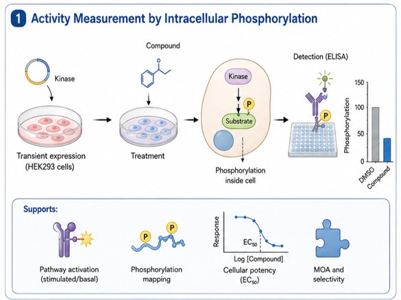
Activity Measurement by Intracellular Phosphorylation
We employ transient expression systems to introduce target kinases into human cell lines, most commonly HEK293-derived backgrounds. Following compound treatment, intracellular phosphorylation levels are quantified using highly specific phospho-antibodies in homogeneous or ELISA-based detection formats. Reduced phosphorylation at the target site provides direct evidence of kinase inhibition and can be normalized to total protein levels for precise quantification. This strategy supports:
Pathway activation studies under stimulated or basal conditions
Intracellular phosphorylation mapping at physiologically relevant sites
Cellular potency (EC50) determination in a native signaling context
Mechanistic studies of compound mode-of-action and selectivity
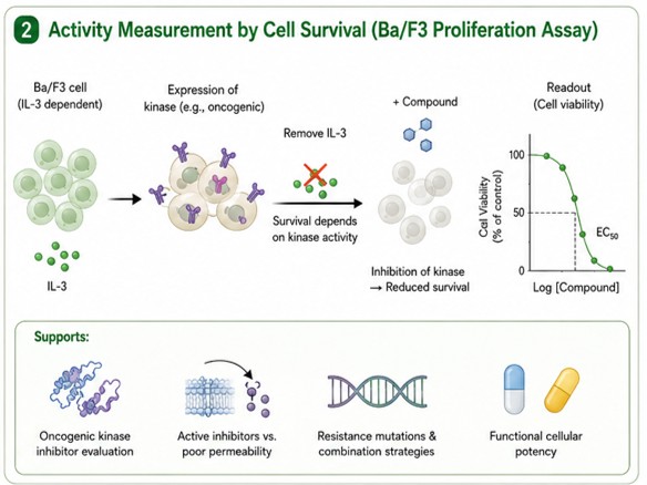
Activity Measurement by Cell Survival (Ba/F3 Proliferation Assay)
Pioneered by Daley and Baltimore (1988), this approach expresses kinases of interest in IL-3–dependent Ba/F3 murine pro-B cells. Upon withdrawal of IL-3, cell proliferation and survival become strictly dependent on the activity of the introduced kinase—either through constitutive activation or downstream signaling rescue. Compound-induced inhibition of the target kinase reduces cell viability, providing a direct, functional readout of cellular potency. This system is particularly powerful for:
Distinguishing active inhibitors from compounds with poor cell permeability
Assessing resistance mutations and combination strategies
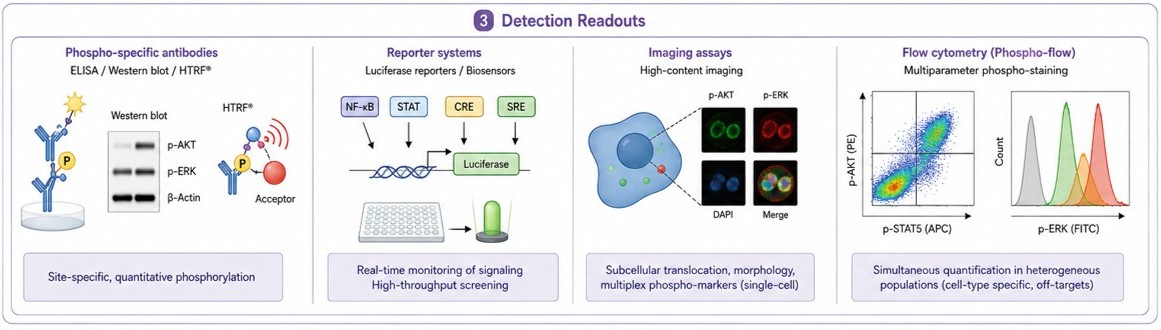
Detection Readouts
Profacgen supports multiple detection modalities to match sensitivity, throughput, and mechanistic requirements:
Phospho-specific antibodies: Sandwich or direct ELISA, western blot, and homogeneous time-resolved FRET using validated antibodies against phospho-epitopes (e.g., phospho-tyrosine, phospho-AKT Ser473, phospho-ERK Thr202/Tyr204). These provide site-specific, quantitative phosphorylation data with high specificity.
Reporter systems: Luciferase-based pathway reporters (e.g., NF-κB, STAT, CRE, SRE) and kinase-specific biosensors enable real-time, high-throughput monitoring of signaling dynamics. Reporter assays are ideal for screening large compound libraries and profiling pathway selectivity.
Imaging assays: High-content imaging platforms quantify subcellular translocation (e.g., AKT membrane recruitment), morphological changes, and multiplexed phospho-marker staining at single-cell resolution. Imaging provides spatial and temporal insights unavailable in population-averaged assays.
Flow cytometry: Phospho-flow cytometry enables simultaneous quantification of kinase pathway activation across heterogeneous cell populations, including primary cells and co-culture systems. Multi-parameter staining distinguishes cell-type–specific responses and off-target effects in complex models.
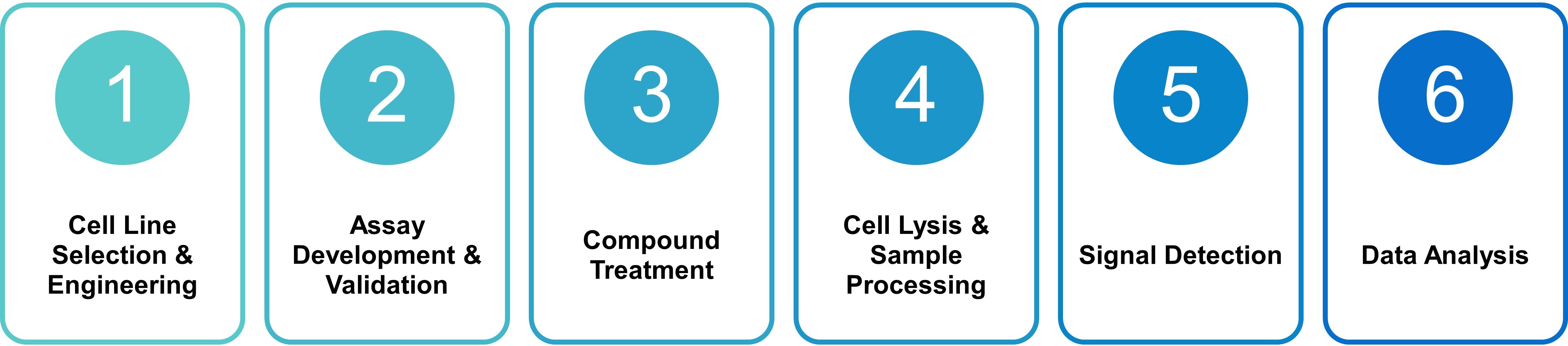
Assay Principle and Workflow
Cell-based kinase assays measure the functional consequence of kinase inhibition within living cells, capturing compound permeability, metabolic stability, and off-target liabilities that biochemical assays miss. Profacgen follows a rigorous, standardized workflow to ensure reproducibility and regulatory compliance:
Cell Line Selection & Engineering: We select or engineer the optimal cell system based on the target kinase and assay format. Options include transient transfection (HEK293), stable knock-in lines, Ba/F3 kinase-dependent proliferation models, and patient-derived or primary cell systems. Cell identity is verified by STR profiling and mycoplasma testing.
Assay Development & Validation: For each cell system, we optimize cell density, stimulation conditions (e.g., growth factor, serum), compound treatment duration, and lysis parameters. Z′ factors and signal-to-background ratios are determined to confirm assay robustness.
Compound Treatment: Test compounds are prepared as DMSO stock solutions and serially diluted across a concentration range (typically 10-point, half-log dilutions). Cells are treated in 96- or 384-well plates for a defined duration (typically 1–24 hours for phosphorylation assays; 48–72 hours for proliferation assays).
Cell Lysis & Sample Processing: Cells are lysed under conditions that preserve phospho-epitopes. For phospho-antibody formats, lysates are transferred to detection plates or processed for western blot. For proliferation assays, viability reagents are added directly to culture wells.
Signal Detection: Detection is performed according to the selected readout—luminescence for viability, fluorescence or TR-FRET for phospho-antibody assays, flow cytometry for phospho-flow, or automated imaging for high-content platforms.
Data Analysis: Raw signals are normalized to vehicle (DMSO) and positive control (reference inhibitor) wells. Percent inhibition or percent viability is calculated and fitted to a four-parameter logistic model to derive EC50 or IC50 values. For mechanistic studies, pathway activation kinetics and combination indices are computed.
Service Capabilities
Capability
Description
Typical Deliverable
Cellular Potency Profiling
EC50 / IC50 determination for lead compounds against target kinases in relevant cell lines, bridging biochemical potency to cellular efficacy.
Dose–response curves; EC50 / IC50 values; Hill slope
Pathway Activation Mapping
Quantification of upstream and downstream signaling node phosphorylation (e.g., RTK → AKT → mTOR → S6K) to assess pathway engagement and feedback loops.
Pathway activation heat map; kinetic profiles
Intracellular Phosphorylation Analysis
Site-specific phospho-antibody detection at native epitopes to confirm on-target inhibition and rule out assay artifacts.
Phospho-site quantification; total protein normalization
Selectivity & Off-Target Profiling
Parallel screening against a panel of cell lines expressing related kinases to evaluate compound selectivity in a cellular context.
Selectivity matrix; fold-shift vs. biochemical data
Mechanism-of-Action Studies
Time-course inhibition, washout recovery, and combination assays to classify reversible vs. irreversible inhibition and synergy.
Reversibility classification; combination indices (CI)
Resistance Mutation Assessment
Evaluation of compound activity against clinically relevant gatekeeper, activation loop, and compound-binding pocket mutations in Ba/F3 or engineered lines.
Fold-shift in EC50; resistance profile summary
Custom Assay Development
Development and validation of novel cell-based assays for non-standard kinases, primary cell models, or co-culture systems.
Qualified protocol; validation report; SOP
Supported Kinase Types
We proudly offer a large selection of kinase targets in cell-based assays:
Tyrosine Kinases
ABL (BCR-ABL)
EphA1
FGFR3
HER2 (ERBB2)
PDGFRβ
ALK
EphA3
FGFR3 [K650M]
HER3 (ERBB3)
RET
ARG (ABL2)
EphA4
FGFR3/BAIAP2L1
IGF1R
RET [V804M]
AXL
EphA5
FGFR4
INSR
RON (MST1R)
BLK
EphB1
FGFR4 [V550E]
JAK1
ROR1
BMX
EphB2
FGR
JAK2
ROS (ROS1)
BTK
EphB4
FLT1 (VEGFR1)
JAK3
RYK
CCK4 (PTK7)
FAK
FLT3
KDR (VEGFR2)
SRC
DDR2
FGFR1
FLT3-ITD
KIT
SYK
EGFR
FGFR1 [V561M]
FLT3-ITD [D835V]
KIT [D816V]
TIE1
EGFR [D746-750]
FGFR2
FLT3-ITD [D835Y]
KIT [K642E]
TIE2
EGFR
[D746-750+T790M]
FGFR2 [K660E]
FLT3-ITD [F691L]
KIT [N822H]
TRKA (NTRK1)
EGFR [L858R]
FGFR2 [K660N]
FLT3-ITD [Y842C]
KIT [T670I]
TRKB (NTRK2)
EGFR
[L858R+T790M]
FGFR2 [N550K]
FLT3-ITD [Y842H]
KIT [V654A]
TRKC (NTRK3)
EGFR [L858R+C797S]
FGFR2 [V565I]
FLT4 (VEGFR3)
LCK
TYK2
EGFR
[L858R+T790M+C797S]
FGFR2/AFF3
FMS (CSF1R)
LYN
TYRO3
EGFR [L861Q]
FGFR2/BICC1
FRK
MER (MERTK)
ZAP70
Serine and Threonine Kinases
AKT1
DCLK2
DYRK1A
MST1
PDK1
PIM1
PIM2
PIM3
Applications
Cell-based kinase assays are essential at multiple stages of the drug discovery and development pipeline:
Kinase inhibitor screening: Primary and secondary screening of small-molecule libraries, natural products, and biologics to identify inhibitors with demonstrable cellular activity, eliminating compounds with poor permeability or metabolic instability.
Cellular potency determination: Quantitative EC50 / IC50 ranking to prioritize leads and establish structure–activity relationships (SAR) that account for cellular context.
Pathway activation and intracellular phosphorylation studies: Mapping of compound effects on complete signaling cascades to confirm on-target mechanism, identify compensatory pathway activation, and guide combination therapy design.
Mechanism-of-action and selectivity profiling: Differentiation of ATP-competitive, allosteric, and covalent inhibitors through cellular kinetics, reversibility, and multi-target profiling.
Resistance and mutant profiling: Preclinical assessment of compound susceptibility to clinically observed resistance mutations, supporting proactive medicinal chemistry and clinical trial design.
Translational pharmacology: Correlation of cellular potency with patient-derived models and biomarker strategies to predict clinical efficacy and dosing regimens.
Service Advantages
Physiologically Relevant Context: Assays performed in intact cells capture permeability, metabolism, and endogenous cofactor effects absent in purified enzyme systems
Multiple Readout Modalities: Phospho-specific antibodies, reporter systems, imaging assays, and flow cytometry to match project needs
Extensive Kinase Panel: Broad coverage of tyrosine and serine/threonine kinases, including wild-type and clinically relevant mutants
Ba/F3 Proliferation Platform: Robust, kinase-dependent survival assay for direct cellular potency and resistance mutation evaluation
High-Throughput Compatibility: 96- and 384-well formats with automated liquid handling for efficient screening
Integrated Data Analysis: Automated curve fitting, pathway mapping, and statistical reporting to accelerate decision-making
Deliverables
Upon project completion, clients receive a comprehensive data package including:
Raw data: Unprocessed plate reader outputs, flow cytometry FCS files, or imaging datasets in standardized formats
Dose–response curves: Graphical plots with fitted four-parameter logistic curves, including 95% confidence intervals and replicate statistics
EC50 / IC50 values: Tabulated potency parameters with cell line, assay format, and treatment conditions
Pathway activation profiles: Phospho-site quantification and signaling node mapping for mechanistic studies
Study report: Detailed protocol description, quality control metrics, statistical analysis, and interpretive summary prepared by our scientific team
Frequently Asked Questions (FAQs)
Q: What is the key difference between cell-based and in vitro kinase assays?
A: In vitro assays use purified kinases in artificial buffer systems and measure direct enzyme inhibition, providing precise biochemical potency (IC50). Cell-based assays evaluate compound activity within living cells, capturing membrane permeability, intracellular ATP competition, metabolic stability, and off-target liabilities. Cell-based EC50 values are generally more predictive of in vivo efficacy and are essential for validating that biochemical hits translate into functional cellular inhibition.
Q: How do you measure intracellular phosphorylation?
A: We quantify intracellular phosphorylation using validated phospho-specific antibodies in HTRF, ELISA, western blot, or flow cytometry formats. Cells are lysed under phosphatase-inhibiting conditions, and phosphorylated epitopes are detected with high specificity. Signals are normalized to total protein or a housekeeping control to ensure accurate, quantitative results.
Q: What cell lines are used for the Ba/F3 proliferation assay?
A: Ba/F3 is an IL-3–dependent murine pro-B cell line originally developed for hematopoietic research. We engineer Ba/F3 cells to express the target kinase (wild-type or mutant) such that kinase activity drives survival and proliferation in the absence of IL-3. This system is widely accepted for evaluating oncogenic kinase inhibitors, including clinically validated targets such as BCR-ABL, FLT3-ITD, and EGFR mutants.
Q: Can you assess compound selectivity in cell-based assays?
A: Yes. We maintain a panel of cell lines expressing related kinases within the same family (e.g., EGFR, HER2, HER4; JAK1, JAK2, JAK3, TYK2). Compounds are screened in parallel to generate a cellular selectivity profile. This approach identifies off-target liabilities that may not be apparent in biochemical assays due to differences in cellular context and compound access.
Q: How long does a typical cell-based kinase assay project take?
A: Standard cellular potency determinations (EC50) require 3–4 weeks, including cell culture, compound treatment, and data analysis. Custom assay development or primary screening campaigns may extend to 6–8 weeks. We provide detailed project timelines during consultation and offer expedited processing for time-critical programs.
Q: Can you evaluate resistance mutations in cell-based assays?
A: Absolutely. Our Ba/F3 platform supports expression of clinically observed resistance mutations (e.g., EGFR T790M, ABL T315I, FLT3 D835V). We compare compound potency against wild-type and mutant constructs to calculate fold-shift values, providing critical data for resistance mitigation strategies and next-generation inhibitor design.
For more information about our cell-based kinase assays, please contact us for details. Our tech representatives are available to help you 24 hours a day, Monday through Friday.
References:
Daley GQ, Baltimore D. Transformation of an interleukin 3-dependent hematopoietic cell line by the chronic myelogenous leukemia-specific P210 bcr/abl protein. Proc Natl Acad Sci USA. 1988;85(23):9312–9316. doi:10.1073/pnas.85.23.9312
Online Inquiry
Fill out this form and one of our experts will respond to you within one business day.