
miCLIP-seq (m6A individual-nucleotide-resolution cross-linking and immunoprecipitation) is a specialized variant of the iCLIP technology designed to detect m6A modifications at single-nucleotide resolution across the transcriptome. Developed by Linder et al. in 2015, miCLIP builds upon the ultraviolet (UV) crosslinking and immunoprecipitation framework by introducing two critical modifications: the use of a highly specific m6A antibody for immunoprecipitation, and the exploitation of characteristic UV-induced mutations at m6A sites to distinguish modified adenosines from their unmodified counterparts at single-base precision. This approach overcomes the resolution limitation of conventional m6A mapping methods such as MeRIP-seq, which identify methylated regions at 100–200 nucleotide resolution but cannot pinpoint individual m6A sites.
N6-methyladenosine (m6A) is the most prevalent internal modification on eukaryotic mRNA, present at an estimated 3–5 sites per transcript, and it plays crucial regulatory roles in virtually every aspect of mRNA metabolism: nuclear export, translation efficiency, splicing, stability, and decay. The m6A modification is deposited co-transcriptionally by the METTL3/METTL14 writer complex, recognized by YTH-domain reader proteins, and can be removed by FTO and ALKBH5 demethylases. Precise knowledge of where m6A is deposited—and how these positions change under different biological conditions—is essential for understanding epitranscriptomic regulation in development, cancer, neurobiology, and viral infection. Profacgen provides a complete miCLIP-seq service for accurate, single-nucleotide-resolution m6A profiling.
Although chemical modifications on RNA were first described in the 1970s, the field of epitranscriptomics—the study of post-transcriptional RNA modifications and their functional consequences—remained dormant for decades due to a lack of methods for precise, transcriptome-wide mapping. The turning point came in 2012, when two independent laboratories reported the first transcriptome-wide m6A maps in human and mouse cells using MeRIP-seq (also called m6A-seq). These studies revealed that m6A is enriched near stop codons, in 3′ UTRs, and within long internal exons, and identified the METTL3/METTL14/WTAP writer complex responsible for m6A deposition.
The subsequent identification of m6A erasers (FTO, ALKBH5) and readers (YTHDF1–3, YTHDC1–2) established m6A as a dynamic, reversible modification analogous to DNA methylation and histone modifications. Functional studies demonstrated that m6A controls stem cell pluripotency, circadian rhythms, immune responses, and cancer progression. However, the ~200 nt resolution of MeRIP-seq proved insufficient for understanding how m6A at specific positions regulates individual mRNAs, or how mutations that create or destroy m6A sites alter gene expression.
miCLIP-seq addressed this critical gap by achieving true single-nucleotide resolution. The method relies on the observation that UV crosslinking of the m6A antibody to its target induces characteristic mutation patterns—primarily C-to-T transitions at the crosslinked adenosine—that serve as a molecular signature of m6A presence. By counting these mutation events across the transcriptome, miCLIP not only identifies m6A sites with base-pair precision but also estimates their modification stoichiometry (the fraction of transcripts carrying m6A at each site). This quantitative, single-base resolution has transformed our understanding of m6A heterogeneity and dynamics.
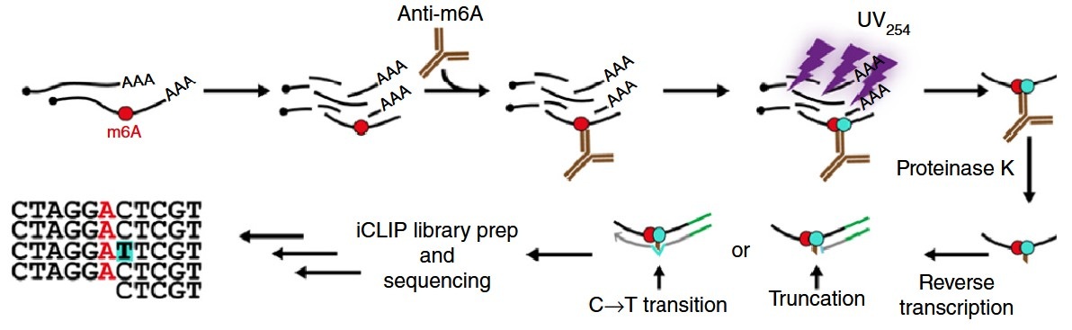 Figure 1. The miCLIP protocol. (Linder et al., 2015)

| Step | Description | Key Parameters |
|---|---|---|
| 1. Sample Preparation | Total RNA or poly(A)+ RNA is extracted and fragmented to 50–150 nt fragments using divalent cation-mediated hydrolysis. mRNA purification removes ribosomal RNA contamination. | RIN ≥ 8; rRNA depletion >95%; Fragment size: 50–150 nt |
| 2. Immunoprecipitation | Anti-m6A antibody conjugated to magnetic beads captures m6A-containing RNA fragments. Pre-clearing with control beads reduces non-specific binding. | Anti-m6A antibody (Synaptic Systems 202-003 or equivalent); 2–4 hr incubation at 4 °C |
| 3. UV Crosslinking | Antibody-RNA complexes are irradiated with 254 nm UV light to create covalent bonds between the antibody and bound RNA nucleotides. This locks the interaction in place for stringent washing. | UV dose: 0.15–0.5 J/cm2; on-bead irradiation |
| 4. Stringent Purification | Crosslinked complexes are washed under high-stringency conditions (SDS, urea, high salt), separated by SDS-PAGE, and transferred to nitrocellulose membrane. Autoradiography (32P-labeled RNA) visualizes the correct size region for excision. | SDS-PAGE: 4–12% Bis-Tris gel; Membrane: 0.45 μm nitrocellulose |
| 5. RNA Recovery & Library Preparation | RNA is released by proteinase K digestion, purified, and subjected to adapter ligation, reverse transcription, and PCR amplification. The resulting cDNA library is size-selected and sequenced on an Illumina platform. | Proteinase K: 200 μg/mL, 1 hr at 37 °C; Library insert: 150–300 bp |
| 6. Bioinformatic Analysis | Sequencing reads are mapped to the reference transcriptome, and m6A sites are identified by the characteristic C-to-T mutation signature using specialized algorithms (miCLIP/MAZTER/m6Atrap). | C-to-T mutation rate threshold: ≥10% above background; FDR < 0.05 |
Single-Nucleotide Resolution
Unlike MeRIP-seq, which maps broad regions of ~100–200 nucleotides, miCLIP precisely identifies the exact adenosine residue carrying the m6A modification, enabling study of site-specific m6A function.
No Nucleotide Pretreatment Required
MiCLIP does not require chemical or enzymatic pretreatment of RNA (unlike PA-m6A-seq or MazF-based methods), preserving the native modification landscape and simplifying the workflow.
Accurate Prediction via Mutation Signature
The characteristic UV-induced mutation pattern at m6A sites (primarily C-to-T transitions) serves as an intrinsic quality control, distinguishing true modifications from technical artifacts with high confidence.
Unbiased by Peak Shape
Because miCLIP identifies m6A at the nucleotide level rather than by peak calling, it is not affected by variable peak shapes, antibody batch effects, or local sequence biases that confound region-based methods.
Stoichiometry Estimation
The frequency of mutation events at each site provides a relative measure of modification stoichiometry—the fraction of transcripts modified at that position—enabling quantitative comparison across conditions.
Distinguishes m6A and m6Am
MiCLIP can discriminate N6-methyladenosine (m6A) from N6,2′-O-dimethyladenosine (m6Am) at the first transcribed nucleotide based on characteristic differences in crosslinking-induced mutation patterns and genomic position.
| Feature | miCLIP-seq | MeRIP-seq | PA-m6A-seq |
|---|---|---|---|
| Resolution | Single nucleotide | 100–200 nt | Single nucleotide |
| RNA pretreatment | None required | None required | Photoactivatable ribonucleoside labeling |
| Quantification | Relative stoichiometry via mutation frequency | Enrichment ratio (IP vs. input) | Mutation rate-based |
| m6Am detection | Yes (discriminates m6A and m6Am) | No | Limited |
| Cell type compatibility | All (UV crosslinking) | All | Requires nucleoside uptake |
| Input RNA | 50–100 μg total RNA | 100–300 μg total RNA | 100–200 μg total RNA |
Background:
A neuro-oncology team hypothesized that m6A modifications on specific transcripts maintained the stemness of glioblastoma stem cells (GSCs), and that targeting the m6A writer METTL3 could deplete the GSC population. They needed a precise map of m6A sites to identify functionally critical methylation events.
Our Solution:
Profacgen performed miCLIP-seq on GSCs and differentiated glioblastoma cells (n = 3 biological replicates each). Total RNA (80 μg per sample) was processed through the complete miCLIP workflow, and libraries were sequenced on the HiSeq 2500 platform to >30 million reads per sample.
Final Results:
miCLIP identified 9,847 high-confidence m6A sites in GSCs, with 2,341 sites showing >2-fold differential methylation compared to differentiated cells. GSC-specific m6A was strongly enriched on transcripts encoding stemness factors (SOX2, MYC, OLIG2) and DNA repair proteins. Notably, a single m6A site in the SOX2 3′ UTR (A1847) showed 85% modification stoichiometry in GSCs vs. 12% in differentiated cells. METTL3 knockdown abolished this methylation, reduced SOX2 protein by 70%, and impaired GSC self-renewal. The data supported a METTL3-targeted therapy approach currently in preclinical development.
Background:
A virology group studying SARS-CoV-2 RNA biology observed that viral replication efficiency varied dramatically between cell types, and hypothesized that host-mediated m6A modification of viral RNA might regulate viral gene expression and immune evasion.
Our Solution:
Profacgen performed miCLIP-seq on SARS-CoV-2 RNA purified from infected Vero-E6 (primate kidney) and Calu-3 (human lung epithelial) cells at 24 hours post-infection. Viral RNA was enriched by ribosomal RNA depletion and oligo(dT) selection before miCLIP processing.
Final Results:
miCLIP identified 42 m6A sites on the SARS-CoV-2 genome, with 28 sites shared between cell types and 14 cell-type-specific sites. The most highly methylated site (A10312 in ORF1a, 62% stoichiometry in Calu-3) mapped to a region encoding the nsp5 protease. Gene knockdown of METTL14 in Calu-3 cells abolished A10312 methylation, increased viral RNA levels 3-fold, and enhanced IFN-β induction—suggesting that m6A on viral RNA suppresses innate immune recognition. The cell-type-specific methylation pattern provided a molecular explanation for the differential viral replication observed between lung and kidney cell lines.
References:
Fill out this form and one of our experts will respond to you within one business day.