We use cookies to understand how you use our site and to improve the overall user experience. This includes personalizing content and advertising. Read our
Privacy Policy
At Profacgen, our Custom Protein Ubiquitination Services deliver comprehensive in vitro and cell-based ubiquitination analysis, site mapping, and quantitative proteomics to support mechanistic studies, degrader validation, and drug discovery programs.
Ubiquitination is one of the most prevalent post-translational modifications in cells. Ubiquitin modification induces protein degradation and affects protein function, localization, and activity. Study of protein ubiquitination therefore plays a critical role in cell biology and drug discovery. As a professional protein degradation services provider, Profacgen offers diverse ubiquitination services to accelerate research progress and generate actionable data.
Overview of Ubiquitination
Ubiquitination is a highly regulated enzymatic cascade that controls protein fate across virtually all cellular processes:
E1: Ubiquitin-activating enzymes catalyze ATP-dependent adenylation and thioester bond formation with ubiquitin, initiating the conjugation cascade. E1 enzymes determine the cellular pool of activated ubiquitin available for downstream transfer
E2: Ubiquitin-conjugating enzymes receive activated ubiquitin from E1 and interact with E3 ligases to facilitate substrate-specific transfer. E2 enzymes influence ubiquitin chain topology, including K48-linked chains for proteasomal degradation and K63-linked chains for signaling
E3: Ubiquitin ligases confer substrate specificity by recognizing degrons, post-translational modifications, or adapter-mediated interactions. E3 ligases are the regulatory hub of ubiquitination and the primary therapeutic target for targeted protein degradation
Polyubiquitination: Sequential ubiquitin conjugation generates chains with distinct linkages (K6, K11, K27, K29, K33, K48, K63, M1) that direct divergent cellular outcomes. Chain length, branching, and mixed topology further expand the ubiquitin code
Figure 1. Schematic representation of the ubiquitination workflow.
Our Ubiquitination Services
Profacgen provides specialized ubiquitination analysis modules tailored to diverse research questions and program stages:
In Vitro Ubiquitination
Reconstituted biochemical assays for precise mechanistic interrogation.
Conjugation reactions: Reconstituted E1-E2-E3 cascades with purified components to determine whether a protein is ubiquitinated, and whether modification is mono-, multi-mono-, or poly-ubiquitin
E2/E3 profiling: Identification of specific E2 conjugating enzymes and E3 ligases responsible for target ubiquitination
Chain topology: Assessment of ubiquitin linkage types by linkage-specific antibodies or mass spectrometry
Cell-Based Ubiquitination
Physiological assessment of ubiquitination in intact cellular environments.
BRET assays: Bioluminescence resonance energy transfer methods to study dynamic ubiquitination of target proteins in living cells in real time
Immunoprecipitation: Co-immunoprecipitation of ubiquitinated substrates with ubiquitin-specific antibodies or TUBE reagents
Pathway modulation: Assessment of ubiquitination changes following degrader treatment, genetic perturbation, or stress induction
Ubiquitination Site Mapping
Precision identification of modified lysine residues.
Di-glycine remnant profiling: Mass spectrometry detection of the Lys-ε-Gly-Gly signature left after tryptic digestion of ubiquitinated proteins
Site-directed mutagenesis: Functional validation of mapped sites by lysine-to-arginine substitution and activity assessment
Quantitative comparison: Relative site occupancy across conditions to identify regulatory hotspots
Ubiquitination Quantification
Accurate measurement of ubiquitin incorporation levels.
Absolute quantification: Stable isotope-labeled ubiquitin standards for precise stoichiometry determination
Relative quantification: SILAC, TMT, or label-free proteomics for comparative ubiquitinome analysis across experimental conditions
Turnover kinetics: Time-course analysis of ubiquitination dynamics following stimulus or degrader treatment
Detection Technologies
Our platform integrates multiple detection modalities to match sensitivity, throughput, and analytical requirements:
Western blot: Traditional antibody-based detection of ubiquitinated proteins using ubiquitin-specific, linkage-specific, or substrate-specific antibodies. Suitable for targeted validation, semi-quantitative assessment, and chain topology analysis
Mass spectrometry: Comprehensive identification of ubiquitination sites, chain linkages, and quantitative ubiquitinome profiling. Our workflow encompasses enzyme digestion, peptide enrichment, LC-MS/MS analysis, and bioinformatics processing to deliver detailed, comprehensive results
ELISA: Quantitative measurement of total ubiquitin and ubiquitinated protein levels in cell lysates, tissue homogenates, and biological fluids. Facilitates high-throughput screening and comparative quantification across large sample sets
Applications
Our ubiquitination services support diverse research and drug discovery applications:
Degrader mechanism validation: Confirmation of target ubiquitination following PROTAC or molecular glue treatment, with assessment of chain topology, site specificity, and proteasome dependence
E3 ligase characterization: Determination of substrate repertoire, ubiquitination kinetics, and chain linkage preferences for novel or disease-relevant E3 ligases
Drug target assessment: Evaluation of compound effects on ubiquitin signaling pathways, including off-target ubiquitination and pathway modulation
Disease mechanism studies: Identification of dysregulated ubiquitination events in disease models, biomarker discovery, and therapeutic target validation
Rich and Comprehensive Services: In vitro reconstitution, live-cell BRET imaging, site-specific mapping, and global proteomics within a single integrated platform.
Professional Evaluation and Experimental Design: Expert consultation to tailor assay selection, experimental conditions, and analysis strategy to your specific biological question.
Stable Platform and Accurate Data: Rigorous assay validation, standardized reagents, and robust quality control ensure reproducible, publication-quality results.
Timely Feedback on Project Progress: Regular milestone updates, transparent communication, and rapid troubleshooting to maintain program timelines.
Representative Program Scenarios
Scenario 1: PROTAC-Induced Ubiquitination Site Mapping
Program Context:
A PROTAC program required precise mapping of ubiquitination sites on a target kinase to confirm mechanism-of-action, optimize degrader design, and predict resistance liabilities.
Objective:
To identify all PROTAC-induced ubiquitination sites, determine chain linkage types, and assess site occupancy relative to basal ubiquitination levels.
Approach:
Profacgen treated target-expressing cells with the PROTAC or vehicle control, then enriched ubiquitinated proteins by TUBE pulldown. Tryptic peptides were analyzed by LC-MS/MS with di-glycine remnant profiling. Site-specific ubiquitination was quantified by label-free quantification, and chain topology was assessed by linkage-specific antibodies. Key sites were validated by lysine-to-arginine mutagenesis and functional degradation assays.
Outcome:
The analysis identified 12 PROTAC-induced ubiquitination sites, with 3 sites showing >5-fold occupancy increase. K48-linked chains predominated, consistent with proteasomal degradation. Mutagenesis of the top site reduced degradation efficiency by 60%, confirming its functional importance and informing degrader optimization to enhance engagement at this critical residue.
Scenario 2: Real-Time BRET Monitoring of Dynamic Ubiquitination
Program Context:
A molecular glue program required kinetic analysis of target ubiquitination to understand the temporal relationship between compound exposure, ubiquitin conjugation, and protein degradation.
Objective:
To establish a live-cell assay for real-time ubiquitination monitoring and correlate kinetics with downstream degradation and cellular phenotypic responses.
Approach:
Profacgen developed a BRET-based ubiquitination sensor by fusing a luciferase reporter to the target protein and a BRET acceptor to ubiquitin. Cells stably expressing the sensor were treated with molecular glue, and BRET signals were recorded in real time over 24 hours. Parallel samples were collected for Western blot and mass spectrometry validation. Degradation kinetics and phenotypic responses were monitored by quantitative imaging and viability assays.
Outcome:
The BRET assay revealed rapid ubiquitination detectable within 15 minutes of compound exposure, peaking at 2 hours and preceding detectable protein loss by 1 hour. This temporal resolution enabled precise correlation of ubiquitination with degradation and cell death, supporting a mechanism-based PK/PD model and guiding dosing strategy for in vivo studies.
Q: What is the difference between mono-ubiquitination and poly-ubiquitination?
A: Mono-ubiquitination is the attachment of a single ubiquitin molecule to one lysine residue, often regulating protein localization, activity, or interaction. Poly-ubiquitination is the formation of ubiquitin chains through successive conjugation, typically targeting proteins for proteasomal degradation (K48-linked) or signaling (K63-linked). Our assays distinguish these modification types by linkage-specific antibodies and mass spectrometry.
Q: Can you detect ubiquitination in living cells?
A: Yes. Our BRET-based assays enable real-time monitoring of ubiquitination dynamics in intact cells without fixation or lysis. This preserves physiological context and captures transient events invisible to endpoint assays. BRET sensors can be customized for specific targets and ubiquitin chain types.
Q: How does mass spectrometry identify ubiquitination sites?
A: Trypsin digestion of ubiquitinated proteins leaves a di-glycine remnant (Lys-ε-Gly-Gly) attached to modified lysines. Our LC-MS/MS platform detects this signature mass shift with high confidence, enabling precise site mapping even in complex samples. Quantitative proteomics further measures relative site occupancy across conditions.
Q: Can you quantify ubiquitination levels absolutely?
A: Yes. We employ stable isotope-labeled ubiquitin standards for absolute quantification of ubiquitin incorporation stoichiometry. Relative quantification by SILAC, TMT, or label-free methods is also available for comparative studies across multiple conditions.
Q: What sample types can you analyze?
A: We analyze recombinant proteins, cell lysates, tissue homogenates, and biological fluids. For in vitro assays, purified E1, E2, E3, and substrate components are reconstituted. For cellular studies, we support transient and stable cell lines, primary cells, and patient-derived samples.
Q: How do you validate that observed ubiquitination is functionally relevant?
A: We validate through multiple approaches: site-directed mutagenesis of identified lysines to assess functional impact; proteasome inhibitor co-treatment to confirm degradation dependence; E3 ligase knockdown or knockout to establish enzyme specificity; and correlation with cellular phenotypic outcomes.
References:
Zuo Y, Chong BK, Jiang K, Finley D, Klenerman D, Ye Y. A general in vitro assay for studying enzymatic activities of the ubiquitin system. Biochemistry. 2020;59(7):851-861. doi:10.1021/acs.biochem.9b00602
Online Inquiry
Fill out this form and one of our experts will respond to you within one business day.
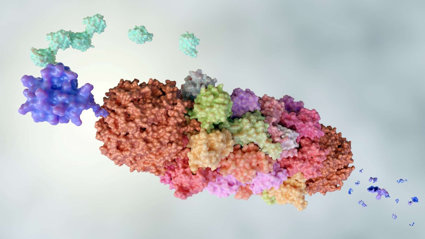
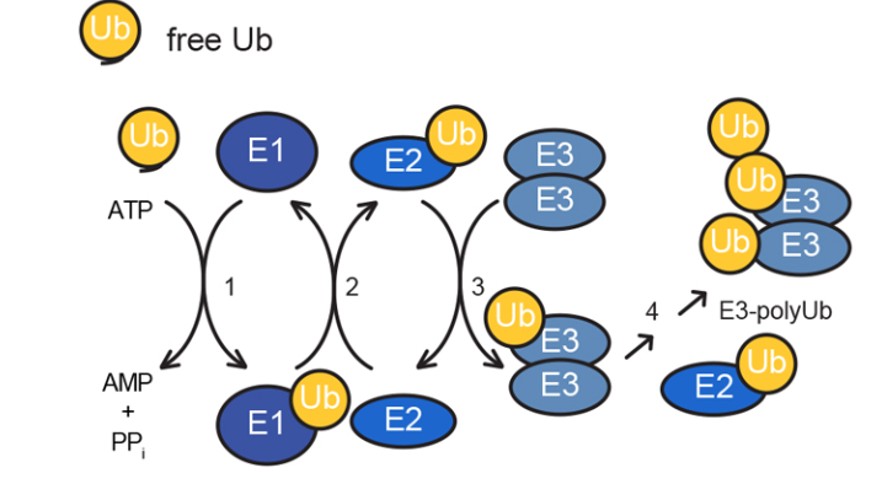 Figure 1. Schematic representation of the ubiquitination workflow.