We use cookies to understand how you use our site and to improve the overall user experience. This includes personalizing content and advertising. Read our
Privacy Policy
Scanning point mutagenesis advances beyond conventional alanine or cysteine scanning by substituting each target amino acid residue with all 20 natural amino acids simultaneously. This powerful technique provides a comprehensive profile of every position within a protein, revealing how side-chain chemistry at each site influences stability, function, binding affinity, and expression. For each codon of interest, a small site-saturation library is constructed, enabling parallel assessment of all possible amino acid alternatives.
Alanine (Ala) scanning remains a widely adopted foundational approach in which residues are systematically replaced with alanine to eliminate side-chain interactions without significantly altering the main-chain conformation or introducing steric and electrostatic perturbations. However, modern protein engineering demands richer information than alanine alone can provide. Profacgen addresses this need through our advanced Shotgun Mutagenesis technology platform, which delivers comprehensive mutagenesis combined with high-throughput phenotypic analysis for any protein of interest.
Background: From Alanine Scanning to Shotgun Mutagenesis
Alanine scanning mutagenesis emerged in the late 1980s as a systematic method for dissecting protein function. The approach rests on a simple but elegant premise: because alanine possesses a methyl side chain that is both small and chemically inert, substituting it for a target residue effectively removes the functional contribution of the original side chain while minimally perturbing the protein backbone. By measuring the functional consequence of each alanine substitution, researchers can identify which positions are critical for activity, binding, or stability.
While transformative, alanine scanning has inherent limitations. By testing only one alternative amino acid at each position, it captures only loss-of-function information—revealing which positions require a side chain, but not which alternative chemistries might enhance function. In the late 1990s, the Weiss laboratory at the University of California introduced combinatorial alanine scanning, which combined alanine substitutions with selected alternative amino acids at key positions, providing richer information than pure alanine scanning.
The field reached its modern form with the development of shotgun scanning mutagenesis, in which every position is replaced with all 20 natural amino acids simultaneously. This comprehensive approach, pioneered by the Doranz laboratory for high-throughput epitope mapping, enables discovery of both loss-of-function and gain-of-function mutations in a single experiment. Profacgen's platform builds on these methodological foundations, integrating automated gene synthesis, high-throughput expression, and quantitative phenotypic screening to deliver complete structure-function maps at single-residue resolution.
Figure 1. Alanine scanning and shotgun scanning mutagenesis. (Adapted from Drici, 2001; and Li et al., 2021)
Conventional alanine scanning identifies specific amino acid residues responsible for peptide stability, function, and conformation by sequentially substituting each non-alanine residue with alanine and measuring the resulting change in activity. While informative, this approach captures only a subset of the chemical diversity available at each position—it tests removal of side-chain functionality but never evaluates alternative chemistries that might enhance or alter activity.
Shotgun scanning mutagenesis overcomes this limitation by replacing every residue with all possible amino acids. This comprehensive substitution matrix enables:
Complete functional mapping: Every side-chain chemistry (polar, charged, hydrophobic, aromatic) is evaluated at every position
Discovery of gain-of-function mutations: Identify substitutions that improve stability, affinity, or catalytic rate beyond wild-type levels
Epitope identification at single-residue resolution: Precisely define antibody-binding footprints by testing all amino acid substitutions at each position
Structural constraint analysis: Determine which positions tolerate substitution (surface loops) versus those that do not (core or active-site residues)
Synergistic interaction detection: Identify positions where mutation combinations produce effects greater than the sum of individual changes
Our Scanning Mutagenesis Services
Service Tier
Description
Deliverables
Alanine Scanning Library
Systematic substitution of each non-alanine residue with alanine
Maintenance of main-chain conformation for side-chain contribution assessment
Classic approach for identifying functionally critical residues
Sequence-verified plasmid library; QC report (SDS-PAGE); Activity comparison data (optional)
Comprehensive Shotgun Scanning
Every position mutated to all 20 natural amino acids
Complete sequence-function matrix for the target region
Compatible with any protein class or size
Complete variant library (20 variants per position); Sequencing verification for all clones; High-throughput screening data (optional)
Targeted Region Scanning
Focused scanning of specific domains, loops, or interfaces
Cost-effective alternative to full-length scanning
Ideal for follow-up studies on regions of interest
Domain-focused variant set; Sequence verification; Expression and purification (optional)
High-Throughput Phenotypic Analysis
Automated expression and screening of scanning libraries
Binding, activity, or stability readouts for every variant
Statistical analysis of position sensitivity and tolerance
Phenotypic dataset per variant; Sensitivity heat maps; Statistical summary report
Key residue identification: Rapidly determine which amino acid positions are essential for protein stability, folding, or function
Protein binding site research: Map interaction interfaces at single-residue resolution by evaluating how each substitution affects binding partner affinity
Enzyme function characterization: Quantify the contribution of each residue to catalytic efficiency, substrate specificity, and allosteric regulation
Epitope mapping: Define antibody-binding sites with precision, guiding therapeutic antibody development and intellectual property strategies
Protein engineering: Identify gain-of-function mutations that enhance expression, solubility, thermostability, or ligand affinity
Protein stability profiling: Assess positional tolerance to substitution, distinguishing structurally constrained regions from flexible, permissive loops
Allosteric site discovery: Identify remote positions where substitution modulates activity at the active site, revealing hidden allosteric networks
Why Choose Profacgen?
Comprehensive Coverage: Unlike traditional approaches limited to alanine or cysteine, our platform tests all 20 amino acids at every position.
High-Throughput Capability: Automated gene synthesis, cloning, expression, and screening process thousands of variants in parallel.
Experienced Technical Team: Our scientists have executed hundreds of scanning mutagenesis projects across diverse protein classes and applications.
Flexible Project Design: Choose full-length scanning, domain-focused analysis, or alanine-only libraries to match your budget and experimental needs.
Case 1: Comprehensive Epitope Mapping of a Therapeutic Antibody Target
Background:
A biopharmaceutical company required precise epitope mapping for a candidate therapeutic antibody targeting a membrane-bound receptor. Traditional peptide-based epitope mapping had provided only approximate binding regions, and the team needed single-residue resolution to support intellectual property filings and rational antibody optimization.
Our Solution:
Profacgen designed a comprehensive shotgun scanning library encompassing the entire extracellular domain of the target receptor (186 residues). Each position was mutated to all 20 natural amino acids, generating 3,720 unique variants. The library was expressed in mammalian cells, and antibody binding was assessed for every variant using a high-throughput flow cytometry assay.
Final Results:
The analysis identified a compact epitope spanning 14 residues, with 3 positions showing absolute conservation of binding (no substitution tolerated) and 4 positions where specific alternative amino acids actually enhanced affinity. These insights enabled the client to file a detailed epitope claim and guided a subsequent affinity maturation campaign that improved binding by 8-fold.
Case 2: Allosteric Network Mapping in a GPCR for Drug Discovery
Background:
A drug discovery team investigating a G protein-coupled receptor (GPCR) needed to map the allosteric communication network linking the orthosteric ligand-binding pocket to the G-protein coupling interface. Understanding this network was essential for designing bitopic ligands that could simultaneously engage both sites.
Our Solution:
Profacgen executed a two-phase scanning campaign. Phase 1 applied alanine scanning to all 312 residues of the receptor's transmembrane and intracellular domains, measuring G-protein activation efficiency for each mutant. Phase 2 performed full shotgun scanning on 28 positions identified as sensitive in Phase 1, introducing all 20 amino acids and measuring both ligand binding (radioligand displacement) and G-protein activation.
Final Results:
The combined analysis revealed a previously uncharacterized allosteric pathway involving 5 interconnected residues that relay conformational changes from the binding pocket to the G-protein interface. Two of these positions, when mutated to tryptophan, constitutively activated the receptor without ligand—confirming their role as conformational switches. This allosteric map directly informed the design of novel bitopic ligands with improved subtype selectivity.
Q: What is the difference between alanine scanning and shotgun scanning mutagenesis?
A: Alanine scanning substitutes each target residue with alanine only, testing the effect of removing side-chain functionality. Shotgun scanning mutagenesis replaces each residue with all 20 natural amino acids, providing a complete profile of how every possible side-chain chemistry affects protein function. Shotgun scanning discovers both loss-of-function and gain-of-function mutations, while alanine scanning primarily identifies loss-of-function positions.
Q: How many variants are generated in a typical scanning library?
A: For a comprehensive shotgun scanning library, the total variant count equals the number of target positions multiplied by 20 (the number of natural amino acids). For example, scanning a 100-residue protein generates approximately 2,000 variants. Targeted region scanning and alanine scanning libraries are correspondingly smaller. We help you design the optimal library scope during project consultation.
Q: Can you scan membrane proteins or large multidomain proteins?
A: Yes. Our platform accommodates proteins of all classes and sizes. For membrane proteins, we typically focus scanning on extracellular or intracellular domains that are accessible to the binding partners or antibodies of interest. For large multidomain proteins, we recommend targeted region scanning of functionally relevant domains to balance comprehensiveness with project feasibility.
Q: What expression systems do you support for scanning libraries?
A: We support scanning library expression in E. coli, yeast (Pichia pastoris and Saccharomyces cerevisiae), insect cells (BEVS/BacMam), and mammalian cells (HEK293, CHO). The optimal system is selected based on your protein's characteristics and the downstream assay requirements.
Q: Do you provide phenotypic screening in addition to library construction?
A: Yes. Profacgen offers integrated high-throughput phenotypic screening services that pair scanning variant libraries with binding assays, activity measurements, or stability assessments. Screening data is delivered as quantitative results with statistical analysis and visualization, transforming raw variant collections into actionable structure-function insights.
Q: How do you handle positions that are essential for protein folding?
A: Positions that are structurally essential typically show a characteristic signature in scanning data: most or all substitutions abolish expression, solubility, or activity. This information is itself valuable, as it identifies structurally constrained regions. We report these "intolerant" positions explicitly in our analysis, and they serve as negative controls that validate the overall experimental system. For projects where maintaining wild-type function is essential, we can preserve critical positions while scanning surrounding regions.
Q: What is the typical timeline for a scanning mutagenesis project?
A: Timelines depend on library size and services selected. A focused alanine scanning library of 50–100 positions typically takes 4–6 weeks. A comprehensive shotgun scanning library for a 150-residue domain requires 8–12 weeks including construction, expression, and optional screening. We provide detailed timelines during project consultation and offer expedited processing for time-sensitive programs.
References:
Drici N. Alanine scanning mutagenesis in Cys-His boxes of human immunodeficiency virus type 1 Nucleocapsid protein P7: Insight from an in silico investigation. Chemical Data Collections. 2022;38:100828. doi:10.1016/j.cdc.2022.100828
Li M, Zheng X, Shanker S, et al. Shotgun scanning glycomutagenesis: A simple and efficient strategy for constructing and characterizing neoglycoproteins. Proc Natl Acad Sci USA. 2021;118(39):e2107440118. doi:10.1073/pnas.2107440118
Online Inquiry
Fill out this form and one of our experts will respond to you within one business day.

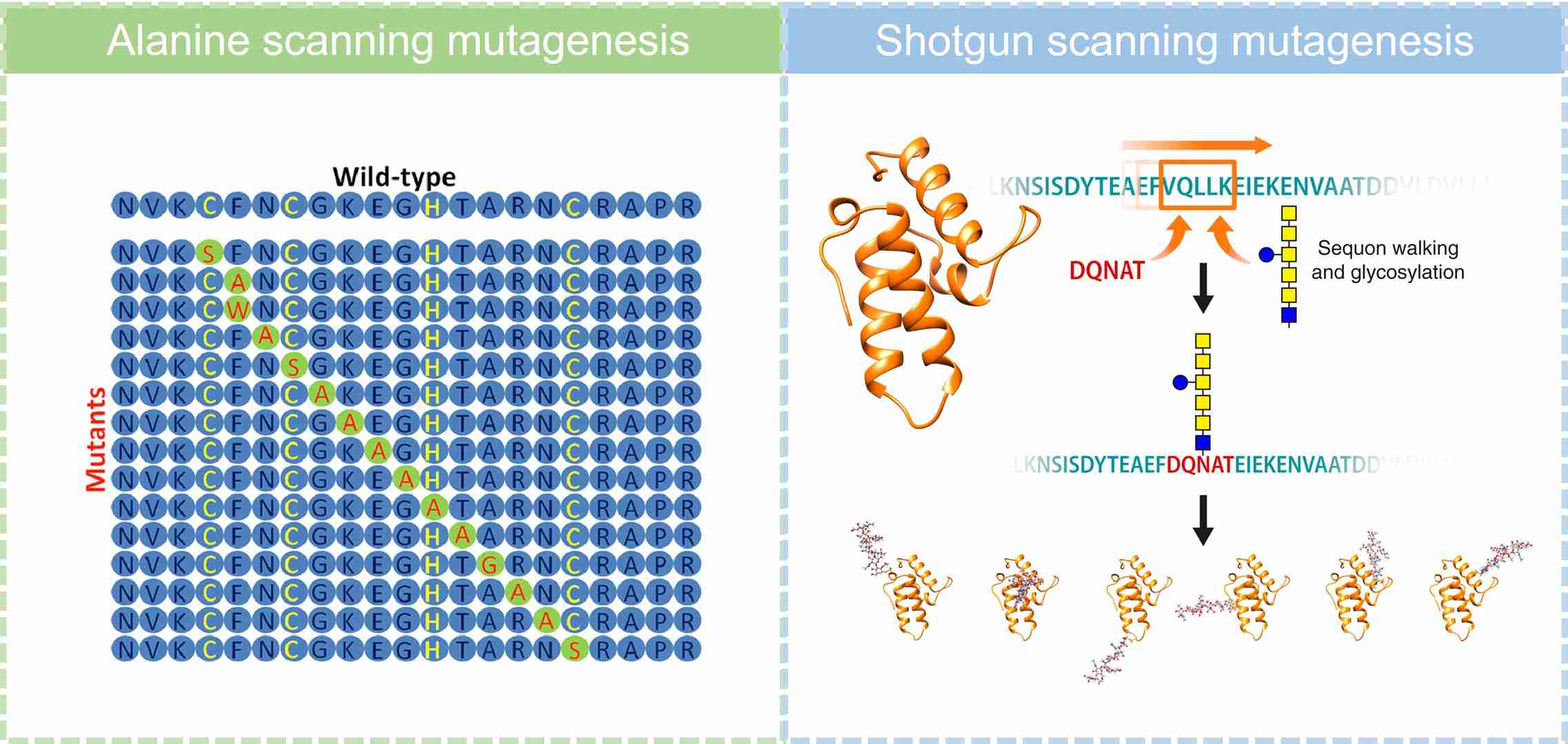 Figure 1. Alanine scanning and shotgun scanning mutagenesis. (Adapted from Drici, 2001; and Li et al., 2021)