
iCLIP-seq (individual-nucleotide resolution UV crosslinking and immunoprecipitation) is a high-resolution method for identifying and precisely mapping protein-RNA interactions across the entire transcriptome. Developed by Jernej Ule and colleagues at the University of Cambridge in 2010, iCLIP overcomes a fundamental limitation of earlier CLIP methods by achieving single-nucleotide resolution in determining where an RNA-binding protein contacts its target RNAs. This precision is essential for understanding the sequence and structural determinants of RNA recognition, the competition between RBPs for overlapping binding sites, and the functional consequences of RNA mutations on protein binding.
The key innovation of iCLIP lies in capturing cDNA truncations that occur during reverse transcription at UV-induced crosslink sites. When a reverse transcriptase encounters a peptide-RNA crosslink, it typically stops one nucleotide before the crosslinked position, leaving a characteristic truncation signature in the cDNA library. By mapping these truncation events to the transcriptome, iCLIP pinpoints the exact nucleotide positions where protein-RNA contacts occur. This resolution is impossible to achieve with standard CLIP methods, which map broad enrichment regions spanning tens to hundreds of nucleotides. Profacgen offers a complete iCLIP-seq service from experimental design through bioinformatic analysis, enabling researchers to explore protein-RNA interactions with unprecedented spatial precision.
Early CLIP methods (HITS-CLIP, PAR-CLIP) revolutionized the study of protein-RNA interactions by enabling genome-wide mapping, but they suffered from limited resolution. HITS-CLIP identified binding regions of approximately 50–200 nucleotides, while PAR-CLIP achieved ~20–50 nucleotide resolution through T-to-C mutation signatures induced by photoactivatable ribonucleoside analogs. While informative, these resolutions were insufficient to discriminate adjacent binding sites within the same regulatory element or to precisely map the RNA sequences recognized by single-strand-specific RBPs.
The breakthrough came from the Ule laboratory's insight that reverse transcriptase stalling at crosslink sites could be harnessed as a positional marker rather than being discarded as a technical artifact. By ligating a 3′ adapter before proteinase K digestion and using an RT primer that introduces a random barcode at the cDNA 5′ end, iCLIP creates a library in which each cDNA molecule records both its truncation position (the crosslink site) and its unique molecular identity (the barcode). This dual encoding enables quantitative analysis of crosslink site distribution and correction for PCR amplification bias.
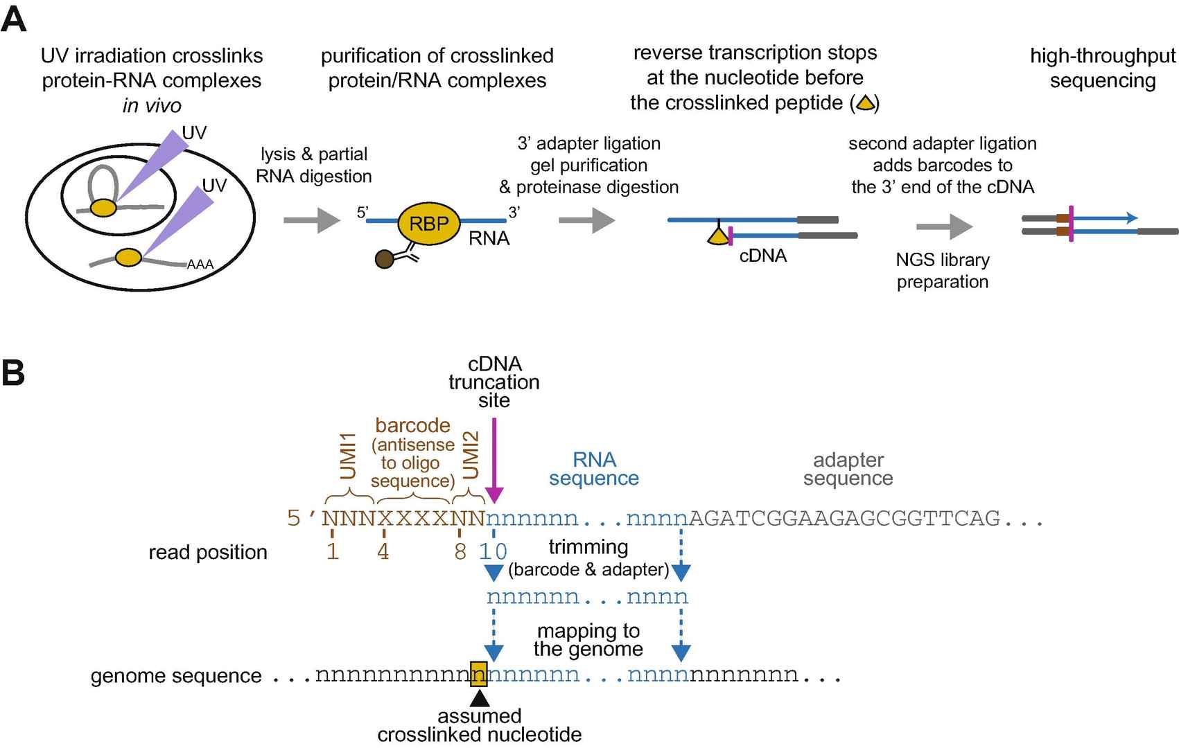 Figure 1. Overview of the iCLIP experiment (A) Experimental workflow. (B) Structure of an iCLIP read. (Busch et al., 2020)
Single-Nucleotide Resolution
cDNA truncation events pinpoint crosslink sites to individual nucleotide positions, revealing the exact protein-RNA contact map across the transcriptome.
UV Crosslinking
254 nm UV irradiation creates zero-length covalent bonds between proteins and directly contacted RNA nucleotides, eliminating non-specific associations and preserving native interaction patterns.
Low Background
Stringent SDS-PAGE purification followed by nitrocellulose membrane transfer effectively removes unbound RNA, un-crosslinked proteins, and non-specific contaminants.
UMI-Based Quantification
Unique molecular identifiers (random barcodes) at the cDNA 5′ end enable PCR deduplication and accurate quantification of crosslink site usage, correcting amplification bias.

| Feature | iCLIP-seq | Standard CLIP-seq | eCLIP-seq |
|---|---|---|---|
| Resolution | Single nucleotide (truncation-based) | 50–200 nt (broad peaks) | 20–50 nt (peak-based) |
| Crosslinking | UV (254 nm) | UV (254 nm) | UV (254 nm) |
| Quantification | UMI-based, PCR-bias corrected | Read-count based | UMI-based, size-matched input |
| Sensitivity | High (detects low-occupancy sites) | Moderate | High (optimized signal-to-noise) |
| Best for | Precise contact site mapping; motif discovery; structural analysis | General binding region identification | Robust peak calling; comparative studies |
Background:
A neurodegeneration research group studying TDP-43, an RNA-binding protein whose cytoplasmic aggregation is the pathological hallmark of amyotrophic lateral sclerosis (ALS) and frontotemporal dementia (FTD), needed a comprehensive map of TDP-43's RNA binding sites to understand how loss of nuclear function contributes to neurodegeneration.
Our Solution:
Profacgen performed iCLIP-seq on TDP-43 in HEK293 cells and human motor neuron-like NSC-34 cells. UV crosslinking, stringent purification, and high-throughput sequencing generated 12.4 million unique cDNA reads. Bioinformatic analysis identified crosslink-induced truncation sites (CITS) using the iCount pipeline, with replicate correlation >0.92.
Final Results:
iCLIP identified 14,687 high-confidence TDP-43 binding sites across 6,241 transcripts, with the UG-rich motif as the dominant binding determinant. Strikingly, 34% of binding sites mapped to intronic regions adjacent to alternatively spliced exons. Comparison with published ALS-associated TDP-43 mutations revealed that 12 mutations directly disrupted iCLIP-identified binding sites, explaining their splicing dysregulation phenotypes. The data provided a molecular rationale for TDP-43 loss-of-function in ALS and identified 8 novel splicing events as potential therapeutic targets.
Background:
An oncology team investigating how the RNA-binding protein HuR stabilizes pro-survival mRNAs under hypoxia required precise binding site information to design decoy oligonucleotides that would competitively block HuR binding and sensitize hypoxic tumor cells to chemotherapy.
Our Solution:
Profacgen performed iCLIP-seq for HuR in MCF-7 breast cancer cells under normoxia (21% O2) and hypoxia (1% O2). Paired-end sequencing on the HiSeq 4000 platform generated >20 million reads per condition. Differential binding analysis was performed using DEseq2 with truncation site counts as input.
Final Results:
HuR binding increased 2-fold at 1,247 sites under hypoxia, with 78% mapping to 3′ UTRs of mRNAs encoding anti-apoptotic proteins (BCL2, MCL1, XIAP). The single-nucleotide resolution revealed that HuR bound a U-rich pentamer (UUUUU) embedded within larger AU-rich elements. A 15-nt decoy oligonucleotide designed against the most strongly bound HuR site in the BCL2 3′ UTR reduced BCL2 mRNA half-life by 60% and restored cisplatin sensitivity in hypoxic MCF-7 cells (IC50 shift from 12 μM to 3 μM).
References:
Fill out this form and one of our experts will respond to you within one business day.