We use cookies to understand how you use our site and to improve the overall user experience. This includes personalizing content and advertising. Read our
Privacy Policy
At Profacgen, our E3 Ligase Activity Assay Services deliver quantitative, high-quality biochemical and cellular characterization of E3 ubiquitin ligase activity, supporting degrader discovery, ligase validation, and mechanistic studies across diverse therapeutic programs.
E3 ubiquitin ligases are key enzymes in the ubiquitin-proteasome pathway. Aberrant E3 ligase activity has been implicated in numerous diseases, making them attractive targets for drug discovery. Accurate analysis of E3 ligase activity is therefore essential for both biochemical studies and degrader development efforts. Profacgen offers comprehensive assay platforms from ligase expression through screening and validation, providing one-stop support for protein degrader programs.
Overview
E3 ligase activity assays measure the critical enzymatic function that drives targeted protein degradation. Understanding this activity requires appreciation of the broader ubiquitination cascade:
E1-E2-E3 cascade: Ubiquitin activation by E1 enzymes, conjugation to E2 carrier proteins, and substrate-specific transfer catalyzed by E3 ligases. E3 ligases confer substrate specificity and determine ubiquitin chain topology, making them the regulatory hub of the pathway
Ubiquitin transfer: E3 ligases catalyze isopeptide bond formation between ubiquitin C-terminus and substrate lysine residues. This transfer can generate mono-ubiquitin, multi-mono-ubiquitin, or polyubiquitin chains with distinct linkages that direct divergent cellular fates
Substrate recognition: E3 ligases recognize substrates through degrons, post-translational modifications, or adapter-mediated interactions. Auto-ubiquitination—a characteristic of most E3 ligases—serves as a convenient readout for activity assessment when exogenous substrates are unavailable
Figure 1. The ubiquitin proteasome system and classification of E3 ubiquitin ligases. (Jeong et al., 2025)
Our Assay Platforms
Profacgen provides multiple assay formats tailored to diverse E3 ligases, screening scales, and analytical requirements:
In Vitro Ubiquitination Assays
Biochemical reconstitution of E3 ligase activity with purified components.
Reconstituted cascades: Purified E1, E2, E3, ubiquitin, and substrate in defined reaction conditions with ATP regeneration
Auto-ubiquitination detection: Measurement of E3 self-modification as a universal activity indicator applicable to most ligases
Substrate-specific assays: Quantification of target protein ubiquitination for substrate-adapted E3 ligases
Cell-Based Activity Assays
Physiological assessment of E3 ligase activity in intact cellular environments.
Endogenous activity profiling: Quantification of native E3 ligase activity by immunoprecipitation and substrate turnover analysis
Overexpression systems: Controlled E3 ligase expression to assess activity, localization, and substrate repertoire in relevant cell lines
Degrader response assays: Cellular evaluation of E3 ligase engagement by heterobifunctional degraders
High-Throughput Assays
Scalable formats for compound screening and comparative ligase profiling.
Plate-based formats: 96-well and 384-well assay configurations for screening compound libraries and E3 ligase panels
Multiplexed detection: Parallel assessment of multiple E3 ligases or ubiquitin chain linkages in single experiments
Automation integration: Robotic liquid handling and detection for consistent, reproducible large-scale screening
Quantitative Activity Analysis
Precise kinetic and thermodynamic characterization of E3 ligase function.
Kinetic profiling: Km and kcat determination for E2-E3 pairs, ubiquitin, and substrate under varied conditions
Thermodynamic analysis: Temperature dependence, pH optima, and cofactor requirements for assay standardization
Inhibitor characterization: IC50, Ki, and mechanism-of-action determination for E3 ligase modulators
Detection Technologies
Our platform integrates multiple detection modalities to match sensitivity, throughput, and quantitative requirements:
Western blot: Traditional antibody-based detection of ubiquitinated proteins. E3 ligase auto-ubiquitination is analyzed by specific antibodies following immunoprecipitation, providing qualitative and semi-quantitative assessment of ubiquitin chain formation
ELISA: Quantitative measurement of ubiquitin and ubiquitinated proteins in serum, plasma, tissue homogenates, cell lysates, and culture supernatants. Facilitates in vitro ubiquitylation of known or putative E3 ligases followed by sensitive colorimetric or chemiluminescent detection
TR-FRET: Time-resolved fluorescence resonance energy transfer utilizing lanthanide chemistry for quantitative, high-sensitivity detection of TUBE-bound polyubiquitin chains. Spatial resolution and extended emission lifetime eliminate background fluorescence from buffers, proteins, compounds, and cell lysates, enabling robust assay performance in complex matrices
Mass spectrometry: Precise identification of ubiquitination sites, chain linkages (K48, K63, K11, etc.), and chain length distributions. Supports quantitative proteomics for global ubiquitinome profiling and substrate repertoire mapping
Assay Readouts
Profacgen provides diverse quantitative and qualitative readouts tailored to program objectives:
Ubiquitination level: Quantification of total ubiquitin incorporation, chain length distribution, and linkage-specific polyubiquitin formation by immunodetection or mass spectrometry
Enzyme activity: Reaction rate determination under standard or varied conditions, including Vmax, specific activity, and fold-activation relative to basal states
Kinetic parameters: Michaelis-Menten analysis for E2, ubiquitin, and substrate Km and kcat values to guide assay optimization and inhibitor evaluation
Substrate specificity: Profiling of ubiquitination efficiency across diverse substrate panels to map E3 ligase recognition motifs and degron preferences
Applications
Our E3 ligase activity assays support diverse targeted protein degradation and drug discovery applications:
E3 ligase characterization: Baseline activity determination, catalytic mechanism elucidation, and comparison with orthologs or disease-associated variants
Degrader screening: Assessment of E3 ligase recruitment by heterobifunctional degraders, ternary complex formation efficiency, and competitive displacement profiles
Mechanistic studies: Dissection of ubiquitin chain topology, processivity, and E2-E3 pairing requirements to inform rational degrader design
Drug discovery: Identification and characterization of E3 ligase activators, inhibitors, and molecular glues that modulate ligase activity for therapeutic benefit
Advanced Technology Platforms: Multiple detection modalities including Western blot, ELISA, TR-FRET, and mass spectrometry matched to your sensitivity and throughput requirements.
Strict and Standardized Process: Rigorous assay validation, standardized reagent preparation, and comprehensive controls ensure reproducible, comparable results across experiments.
Professional Team and Extensive Experience: Deep expertise in ubiquitin biology, E3 ligase biochemistry, and assay development across diverse ligase families and therapeutic targets.
Highly Reliable Results and Analysis: Robust statistical frameworks, appropriate replicate design, and expert data interpretation provide confidence for decision-making.
Short Turn-Around Time and Competitive Price: Efficient workflows and scalable platforms deliver rapid results without compromising quality or analytical rigor.
Representative Program Scenarios
Scenario 1: TR-FRET-Based High-Throughput Screening of E3 Ligase Modulators
Program Context:
A drug discovery program sought to identify small molecule activators of a disease-associated E3 ligase to enhance substrate degradation. Traditional Western blot methods were insufficient for screening a 50,000-compound library.
Objective:
To develop and execute a robust, high-throughput E3 ligase activity assay capable of identifying activators with sub-micromolar potency and confirmed mechanism.
Approach:
Profacgen developed a TR-FRET assay utilizing lanthanide donor-acceptor pairs to detect polyubiquitin chain formation by the target E3 ligase. The assay was optimized for Z'-factor, dynamic range, and DMSO tolerance in 384-well format. The compound library was screened at 10 µM with orthogonal confirmation by Western blot and ELISA. Hits were validated by dose-response analysis, kinetic profiling, and substrate-specific ubiquitination assessment.
Outcome:
The screen identified 23 confirmed activators with EC50 values < 1 µM. Lead compounds increased E3 ligase activity by 3–8 fold with no detectable off-target effects against a panel of 20 related ligases. Two candidates progressed to cellular validation, demonstrating enhanced substrate turnover and supporting advancement to in vivo proof-of-concept studies.
Scenario 2: Comparative E3 Ligase Profiling for Degrader Optimization
Program Context:
A PROTAC program required selection of the optimal E3 ligase recruiter from a panel of candidates for a challenging kinase target. The team needed quantitative activity data to predict ternary complex efficiency and cellular degradation potency.
Objective:
To profile the ubiquitination activity of five candidate E3 ligases and correlate catalytic efficiency with degrader performance in cellular models.
Approach:
Profacgen reconstituted in vitro ubiquitination assays for each E3 ligase with matched E2 conjugating enzymes and model substrates. Auto-ubiquitination rates, substrate turnover kinetics, and chain topology were quantified by TR-FRET and mass spectrometry. Ternary complex formation with the target warhead was assessed by SPR. Degrader prototypes recruiting each ligase were synthesized and tested for cellular degradation potency.
Outcome:
Activity profiling revealed a 10-fold range in catalytic efficiency across the candidate panel. The highest-activity E3 ligase correlated with superior cellular degradation potency, validating in vitro activity as a predictive parameter. The optimized degrader achieved sub-nanomolar DC50 with maximal degradation >90%, supporting selection of the high-activity ligase for lead optimization.
Q: What is auto-ubiquitination and why is it used for E3 ligase activity assays?
A: Auto-ubiquitination is the self-modification of E3 ligases with ubiquitin. It is a characteristic of most E3 ligases and serves as a convenient universal readout when specific exogenous substrates are unavailable. We detect auto-ubiquitination by Western blot, ELISA, or TR-FRET to assess ligase catalytic competence.
Q: What is the difference between TUBEs and TR-FRET for ubiquitin detection?
A: TUBEs (Tandem Ubiquitin Binding Entities) are high-affinity reagents that capture polyubiquitin chains for detection. TR-FRET utilizes lanthanide fluorophores to quantitate TUBE binding with exceptional sensitivity and minimal background. TR-FRET is preferred for high-throughput screening, while TUBEs alone are suitable for rapid, cost-effective characterization.
Q: Can you measure activity for E3 ligases without known substrates?
A: Yes. Auto-ubiquitination serves as a substrate-independent activity readout for most E3 ligases. For ligases with minimal auto-ubiquitination, we employ E2 discharge assays or profile candidate substrates from ubiquitinome databases to identify suitable reaction partners.
Q: What is the typical throughput for your E3 ligase assays?
A: Western blot and ELISA formats typically support tens to hundreds of samples per experiment. TR-FRET and automated platforms enable thousands of reactions in 384-well format for high-throughput screening. We match assay format to project scale and analytical requirements.
Q: Can you assess E3 ligase activity in crude cell lysates?
A: Yes. TR-FRET is particularly suited for complex matrices including cell lysates due to its time-resolved detection that eliminates background fluorescence. We also offer immunodepletion and enrichment strategies to enhance signal-to-noise in crude preparations.
Q: How do you validate assay reproducibility?
A: We calculate Z'-factor for high-throughput assays, implement inter-plate and inter-day controls, and report coefficient of variation across replicates. Standard curves and reference compounds are included in every experiment to ensure consistency and enable cross-study comparison.
References:
Jeong Y, Oh AR, Jung YH, Gi H, Kim YU, Kim K. Targeting E3 ubiquitin ligases and their adaptors as a therapeutic strategy for metabolic diseases. Exp Mol Med. 2023;55(10):2097-2104. doi:10.1038/s12276-023-01087-w
Online Inquiry
Fill out this form and one of our experts will respond to you within one business day.

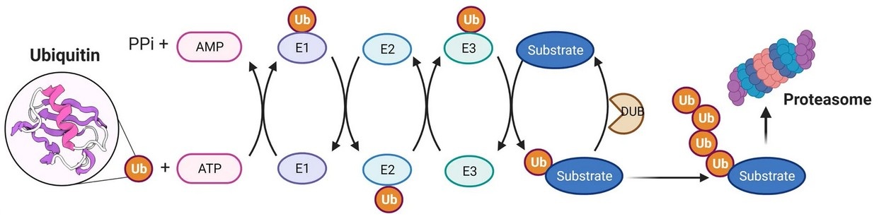 Figure 1. The ubiquitin proteasome system and classification of E3 ubiquitin ligases. (Jeong et al., 2025)