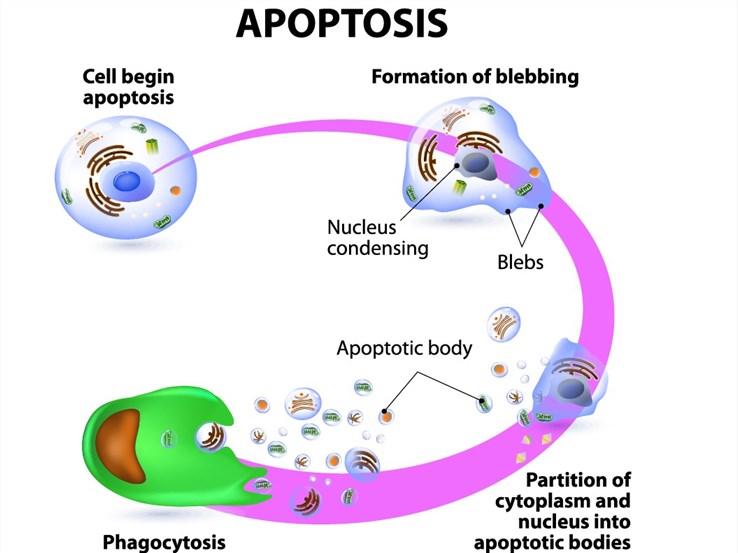
Apoptosis, or programmed cell death, is a highly regulated process that eliminates damaged, infected, or redundant cells without triggering inflammatory responses. Unlike necrosis, apoptosis is characterized by distinct morphological changes including cell shrinkage, chromatin condensation, nuclear fragmentation, plasma membrane blebbing, and the formation of membrane-enclosed apoptotic bodies. These changes result from the activation of conserved proteolytic cascades, primarily involving caspase family proteases, and represent a fundamental mechanism for tissue homeostasis, immune regulation, and embryonic development.
Dysregulation of apoptosis is a hallmark of numerous diseases. The ability of tumor cells to evade apoptotic signaling underlies cancer progression and therapeutic resistance, while excessive apoptosis contributes to neurodegenerative disorders, ischemia-reperfusion injury, and autoimmune pathologies. Consequently, the ability to measure and modulate apoptotic pathways is central to drug discovery, toxicology assessment, and mechanistic studies in both oncology and immunology.
At Profacgen, we provide comprehensive Apoptosis Assay Services designed to detect, quantify, and characterize apoptotic events across all stages of the cell death program. Our platform supports small-molecule screening, biologics characterization, mechanism-of-action studies, and safety assessment for therapeutic candidates targeting apoptotic pathways.
Apoptosis proceeds through a temporally ordered sequence of biochemical and morphological events that provide multiple analytical windows. Understanding these stages is essential for selecting the appropriate detection method:
Figure 1. Overview of intrinsic and extrinsic apoptotic pathways with corresponding assay detection points.
Profacgen offers a comprehensive panel of apoptosis detection methods, each targeting specific biochemical events within the apoptotic program. Assay selection is guided by the research objective, cell type, throughput requirements, and whether early, intermediate, or late apoptotic events are of primary interest.
| Apoptotic Stage | Biomarker/Event | Detection Method | Readout |
|---|---|---|---|
| Early | Phosphatidylserine (PS) externalization | Annexin V binding assay | Flow cytometry or fluorescence microscopy; PS exposure on outer leaflet |
| Early | Mitochondrial membrane potential (MMP) loss | JC-1, TMRE, or DiOC6 staining | Fluorometric or flow cytometric detection of depolarization |
| Intermediate | Caspase activation | Caspase activity assays (fluorogenic or colorimetric substrates) | Substrate cleavage kinetics; specific caspase-3, -8, or -9 activity |
| Intermediate | Caspase substrate cleavage | Western blot or immunofluorescence for PARP, lamin A/C, cytokeratin 18 | Appearance of signature cleavage products |
| Late | DNA fragmentation | TUNEL (terminal deoxynucleotidyl transferase dUTP nick end labeling) | Flow cytometry, microscopy, or plate reader; labeled DNA strand breaks |
| Late | Chromatin condensation | Hoechst 33342 or DAPI staining | Fluorescence microscopy; nuclear morphology assessment |
| General | Live/dead discrimination | Annexin V/PI or Annexin V/7-AAD dual staining | Flow cytometry; quadrant analysis of viable, early apoptotic, late apoptotic, and necrotic populations |
Externalization of phosphatidylserine (PS) from the inner to the outer leaflet of the plasma membrane is one of the earliest detectable events in apoptosis. Annexin V, a 35–36 kDa calcium-dependent phospholipid-binding protein, exhibits high affinity for PS-containing membranes. In viable cells, PS is sequestered on the cytoplasmic face of the membrane; during early apoptosis, caspase-activated scramblase activity and inhibition of aminophospholipid translocase expose PS on the cell surface.
Profacgen offers Annexin V assays in multiple formats:
Loss of mitochondrial membrane potential (ΔΨm) is an early indicator of intrinsic pathway activation. We employ cationic lipophilic dyes including JC-1 (which shifts from red J-aggregates to green monomers upon depolarization), tetramethylrhodamine ethyl ester (TMRE), and 3,3′-dihexyloxacarbocyanine iodide (DiOC6) to monitor ΔΨm changes by flow cytometry or fluorometry.
Caspases are cysteine-aspartic proteases that serve as the central executioners of apoptosis. Caspase-8 and -10 initiate the extrinsic pathway; caspase-9 initiates the intrinsic pathway; and caspase-3, -6, and -7 execute the downstream proteolytic program. Profacgen provides caspase activity assays using fluorogenic or colorimetric tetrapeptide substrates:
Assays can be performed in cell lysates for endpoint measurement or in live cells using cell-permeable substrates for kinetic analysis. We also support caspase substrate cleavage detection by western blot (PARP, lamin A/C) and immunofluorescence for subcellular localization studies.
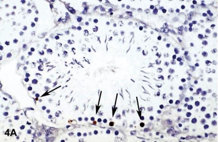
DNA fragmentation is a hallmark of late-stage apoptosis, mediated by caspase-activated DNase (CAD) following ICAD/DFF45 cleavage. The TUNEL (terminal deoxynucleotidyl transferase dUTP nick end labeling) assay detects DNA strand breaks by enzymatically incorporating labeled nucleotides at the 3′-OH ends of fragmented DNA.
Profacgen offers TUNEL assays in multiple configurations:
It is important to note that TUNEL positivity can also occur in necrotic cells or cells undergoing DNA repair. We recommend combining TUNEL with Annexin V staining or caspase activity measurement to confirm apoptotic etiology.
Nuclear morphological changes are assessed using membrane-permeable DNA dyes such as Hoechst 33342 or DAPI. Apoptotic nuclei exhibit characteristic hypercondensation and fragmentation, visible as bright, punctate staining by fluorescence microscopy. This method is particularly valuable for confirmatory imaging in conjunction with biochemical assays.

Challenge:
A pharmaceutical client developed a small-molecule BCL-2 inhibitor for acute myeloid leukemia (AML) and required comprehensive apoptosis profiling to support IND-enabling studies. The client needed to demonstrate that the compound induced apoptosis specifically through the intrinsic mitochondrial pathway, distinguish on-target BCL-2 inhibition from off-target cytotoxicity, and establish a pharmacodynamic biomarker strategy for clinical translation.
Solution:
Profacgen designed a multi-parameter apoptosis profiling panel across three AML cell lines (MV4-11, MOLM-13, OCI-AML3) with varying BCL-2 dependency. We employed a time-course approach measuring mitochondrial membrane potential (JC-1), caspase-9 and -3 activation (fluorogenic substrates), cytochrome c release (cell fractionation and western blot), and DNA fragmentation (TUNEL). To confirm pathway specificity, we included the BCL-2 selective inhibitor venetoclax as a comparator and used BH3 profiling to validate BCL-2 dependency in each cell line.
Outcome:
The compound induced rapid ΔΨm loss (detectable at 4 hours), followed by caspase-9 activation (6 hours), caspase-3 activation (8 hours), and TUNEL positivity (16 hours)—consistent with ordered intrinsic pathway activation. BH3 profiling confirmed that responsive cell lines exhibited BCL-2 dependency, while resistant lines showed MCL-1 or BCL-XL dependence. The data package supported the client's IND submission and informed the selection of caspase-3 cleavage in peripheral blood mononuclear cells as a clinical pharmacodynamic biomarker.
Challenge:
An ADC developer needed to characterize the apoptotic profile of their candidate across target-expressing tumor cells and target-negative normal cells to demonstrate a therapeutic window. The ADC payload was a microtubule inhibitor, and the client sought to confirm that apoptosis induction correlated with target-dependent internalization rather than nonspecific payload release.
Solution:
Profacgen conducted parallel Annexin V/PI flow cytometry assays, caspase-3/7 activity measurements, and PARP cleavage western blots across a panel of target-positive (HER2+ breast cancer, gastric cancer) and target-negative (HER2− fibroblast, hepatocyte) cell lines. Time-course analysis tracked the progression from early apoptosis (Annexin V+/PI−) to late apoptosis (Annexin V+/PI+) at 24, 48, and 72 hours. A non-targeting isotype ADC and free payload were included as controls.
Outcome:
Target-positive cells exhibited dose-dependent apoptosis with an EC50 of 0.8 nM, while target-negative cells showed no significant apoptosis at concentrations up to 100 nM. The temporal progression from PS externalization to caspase activation to PARP cleavage confirmed ordered apoptotic execution. Free payload induced apoptosis in both cell types at 10 nM, confirming that ADC conjugation conferred the desired selectivity. The data supported the ADC's therapeutic index and informed dose-selection for subsequent xenograft studies.
Request a Project Consultation
References:
Fill out this form and one of our experts will respond to you within one business day.