We use cookies to understand how you use our site and to improve the overall user experience. This includes personalizing content and advertising. Read our
Privacy Policy
Enzyme Fragment Complementation Assay (EFCA) is a powerful protein-protein interaction (PPI) detection technology built on a simple but elegant principle: when certain proteins are split into two inactive fragments, neither piece retains enzymatic function. However, when these fragments are brought into close proximity by the interaction of two fused target proteins, they undergo specific non-covalent complementation and reassemble into a fully functional enzyme—restoring catalytic activity that can be detected with extraordinary sensitivity using standard laboratory instrumentation.
EFCA bridges the gap between the high sensitivity of biochemical enzyme assays and the biological relevance of cell-based interaction detection. Unlike methods that require cell lysis or specialized imaging equipment, EFCA produces a quantitative enzymatic signal proportional to interaction strength, enabling both sensitive detection of weak or transient interactions and robust high-throughput screening of interaction libraries and compound collections. Profacgen has developed a versatile EFCA platform employing β-galactosidase, luciferase, and dihydrofolate reductase as reporter enzymes, offering flexible solutions tailored to the sensitivity, dynamic range, and throughput requirements of your project.
Background: From Genetic Screens to Drug Discovery
The conceptual origins of fragment complementation trace back to the 1960s, when geneticists discovered that certain missense mutations in the lacZ gene encoding β-galactosidase could be rescued by intragenic complementation—two inactive mutant proteins regaining function when co-expressed in the same cell. Ullmann and colleagues formalized this observation in 1967, demonstrating that purified β-galactosidase fragments could spontaneously reassemble in vitro into active enzyme. This foundational work established that protein complementation was not merely a genetic curiosity but a predictable physical phenomenon with practical applications.
The transformation of fragment complementation into a general-purpose PPI detection tool occurred in the late 1990s, when two independent laboratories demonstrated that splitting β-galactosidase into an N-terminal α-fragment (enzyme donor, ED) and a C-terminal ω-fragment (enzyme acceptor, EA) and fusing each to potentially interacting proteins created a genetically encodable interaction sensor. If the target proteins interacted, the ED and EA fragments were juxtaposed, complementing to restore β-galactosidase activity detectable by simple colorimetric, fluorometric, or chemiluminescent substrates.
The development of additional reporter systems—firefly and Renilla luciferase, dihydrofolate reductase (DHFR), and β-lactamase—has dramatically expanded the EFCA toolkit. Each reporter offers distinct advantages: β-galactosidase provides robust colorimetric detection suitable for bacterial and mammalian systems; luciferase achieves the highest sensitivity for detecting weak interactions in mammalian cells; and DHFR enables survival-based selection in bacteria, enabling the screening of libraries containing billions of variants. Profacgen's multi-reporter EFCA platform leverages these complementary strengths to match the optimal detection system to your specific experimental question.
Figure 1. Protein-fragment complementation assay strategy based on the enzyme dihydrofolate reductase. (Remy et al., 2017)
Our EFCA Reporter Systems
β-Galactosidase (β-gal) EFCA
The classic complementation system with versatile detection options.
Protein-protein interaction validation: Confirm interactions identified by Y2H, co-IP, or mass spectrometry in a cellular context with quantitative signal output
Interaction network mapping: Systematically test pairwise or higher-order combinations of proteins within a pathway to build comprehensive interaction maps
Protein interaction inhibitor screening: High-throughput identification of small molecules that disrupt specific disease-relevant PPIs by measuring reduced enzymatic signal
Antibody characterization: Quantify the binding strength of antibody-antigen interactions and map epitopes using domain-deletion mutants
Transient interaction detection: Capture short-lived interactions that dissociate too quickly for detection by reversible methods such as FRET
Allosteric regulation studies: Detect ligand-induced conformational changes that alter protein interaction surfaces, revealing allosteric mechanisms
Cellular pathway analysis: Monitor how drug treatment, genetic perturbation, or signaling events modulate protein interaction networks in real time
Why Choose Profacgen?
Multiple Reporter Options: β-galactosidase, firefly/Renilla luciferase, and DHFR systems for maximum flexibility.
Quantitative Signal Output: Continuous enzymatic signal proportional to interaction strength, enabling ranking and kinetic analysis.
All Subcellular Compartments: Detect interactions in the nucleus, cytoplasm, and at the membrane—not limited to nuclear systems.
HTS-Compatible: Plate reader and flow cytometry formats enable screening of thousands to millions of compounds or library variants.
Orthogonal Validation: Co-immunoprecipitation, immunofluorescence, and cellular imaging available to confirm EFCA results.
Representative Case Studies
Case 1: EFCA-Based HTS Identifies First-in-Class Inhibitors of the KEAP1-NRF2 Interaction
Background:
A drug discovery program targeting the KEAP1-NRF2 antioxidant response pathway required a robust cell-based assay to identify compounds that disrupt the KEAP1-NRF2 protein-protein interaction. KEAP1 ubiquitinates NRF2 under basal conditions; disruption stabilizes NRF2, activating cytoprotective gene expression. Existing assays relied on immunoblotting, which was too low-throughput for library screening.
Our Solution:
Profacgen developed a luciferase-based EFCA in HEK293 cells. The KEAP1 Kelch domain was fused to the N-terminal fragment of firefly luciferase, and the NRF2 ETGE motif-containing peptide was fused to the C-terminal fragment. Interaction restored ~5% of full luciferase activity—sufficient for robust detection. The assay was miniaturized to 384-well format (Z′ = 0.74) and used to screen 75,000 structurally diverse compounds.
Final Results:
18 compounds reduced the EFCA signal by >50%. Five were confirmed by orthogonal co-IP and NRF2 luciferase reporter assays. The lead compound, PK-184, disrupted the KEAP1-NRF2 interaction with an IC50 of 180 nM and showed no activity in a panel of 20 unrelated PPIs, confirming specificity. In A549 lung cancer cells, PK-184 increased NRF2 target gene expression (HO-1, NQO1) by 5-fold and suppressed IL-6-induced inflammation. The compound entered IND-enabling studies for COPD and acute lung injury.
Case 2: DHFR-EFCA Maps the Complete Interaction Landscape of a GPCR Signalosome
Background:
An academic research group studying the β2-adrenergic receptor (β2AR) signaling complex sought to systematically map all binary interactions among 18 proteins known to participate in β2AR signaling—including the receptor itself, Gα subunits, Gβγ, β-arrestins, GRKs, and downstream effectors. Co-IP approaches had identified only a subset of interactions and could not distinguish direct binding from bridged complexes.
Our Solution:
Profacgen employed the DHFR-EFCA system in E. coli to test all 153 pairwise combinations. Each protein was fused to both the F[1,2] and F[3] DHFR fragments, and survival on trimethoprim-selective media was scored as a positive interaction. A reciprocal matrix design (testing A-F[1,2] + B-F[3] and A-F[3] + B-F[1,2]) controlled for orientation effects. All putative positives were validated by β-galactosidase EFCA in mammalian cells.
Final Results:
The screen identified 47 direct binary interactions, 23 of which were novel. Notably, GRK2 directly bound both the β2AR C-terminus and Gβγ, suggesting GRK2 serves as a molecular bridge coupling receptor phosphorylation to G-protein desensitization. The complete interaction map revealed a highly interconnected network with β-arrestin2 as the central hub (9 interactions), explaining its multifunctional role in GPCR signaling, trafficking, and scaffolding. The data were deposited in a public interaction database and formed the basis of 3 subsequent publications.
Q: How does EFCA differ from BiFC for protein interaction detection?
A: Both EFCA and BiFC are fragment complementation technologies, but they use different readouts. EFCA restores enzymatic activity (catalytic signal amplification), while BiFC restores fluorescence (direct optical signal). EFCA offers several advantages: (1) enzymatic signal amplification provides higher sensitivity for weak interactions; (2) the readout is quantitative and compatible with standard plate readers, flow cytometry, and survival selection; (3) some EFCA systems (DHFR) enable library screening at unprecedented scale (108–109 variants). BiFC's advantage is subcellular resolution via fluorescence microscopy. The choice depends on whether sensitivity/quantitation (EFCA) or spatial localization (BiFC) is more important for your project.
Q: Which EFCA reporter system should I choose?
A: The optimal reporter depends on your application: Choose β-galactosidase for robust, cost-effective detection in bacteria, yeast, or mammalian cells with multiple substrate options (colorimetric, fluorescent, chemiluminescent). Choose luciferase for maximum sensitivity when detecting weak interactions in mammalian cells, or when working with limited cell numbers. Choose DHFR for ultra-large library screening (>107 variants) where survival-based selection provides the necessary throughput. Our scientists will recommend the best system based on your target proteins, expected interaction strength, and experimental goals.
Q: Can EFCA detect interactions at the cell membrane?
A: Yes. Unlike transcription-based methods such as yeast two-hybrid that are restricted to the nucleus, EFCA functions in any subcellular compartment. Membrane protein interactions can be detected by targeting the ED and EA fusion proteins to the plasma membrane, endoplasmic reticulum, or mitochondria using appropriate signal peptides and transmembrane anchors. We have successfully applied EFCA to study GPCR oligomerization, receptor tyrosine kinase dimerization, and immune receptor signaling at the cell surface.
Q: How do you distinguish true interactions from spontaneous fragment complementation?
A: Spontaneous complementation (background signal from fragments associating without protein interaction) is controlled through several strategies: (1) rigorous negative controls including each fragment expressed alone and non-interacting protein pairs; (2) using low-affinity fragments that only complement efficiently when driven by specific protein interaction; (3) testing both fusion orientations (ED-ProteinA + ProteinB-EA and ProteinA-ED + EA-ProteinB); (4) introducing interaction-disrupting point mutations as specificity controls; and (5) measuring signal in a concentration-dependent manner to confirm interaction-driven saturation behavior.
Q: What is the dynamic range of EFCA signals?
A: The dynamic range varies by reporter system. Luciferase EFCA offers the widest range (>5 orders of magnitude from background to full signal), making it ideal for detecting weak interactions and ranking compounds by potency. β-galactosidase EFCA provides a 2–3 log dynamic range, sufficient for most interaction validation and screening applications. DHFR survival selection is binary (growth/no growth) but can be coupled with quantitative ONPG assays for signal quantification. During assay development, we optimize fragment affinity and expression levels to maximize the signal window for your specific protein pair.
Q: How long does an EFCA project typically take?
A: A standard EFCA project takes 3–5 weeks: plasmid construction and sequence verification (1–2 weeks), expression system setup and pilot testing (1 week), assay optimization and validation (1 week), and data collection and analysis (1 week). High-throughput screening projects add 2–4 weeks depending on library size. Large-scale pairwise interaction mapping projects (e.g., 100+ combinations) require 6–10 weeks. We provide detailed timelines during project consultation.
References:
Remy I, Campbell-Valois FX, Michnick SW. Detection of protein–protein interactions using a simple survival protein-fragment complementation assay based on the enzyme dihydrofolate reductase. Nat Protoc. 2007;2(9):2120-2125. doi:10.1038/nprot.2007.266
Online Inquiry
Fill out this form and one of our experts will respond to you within one business day.

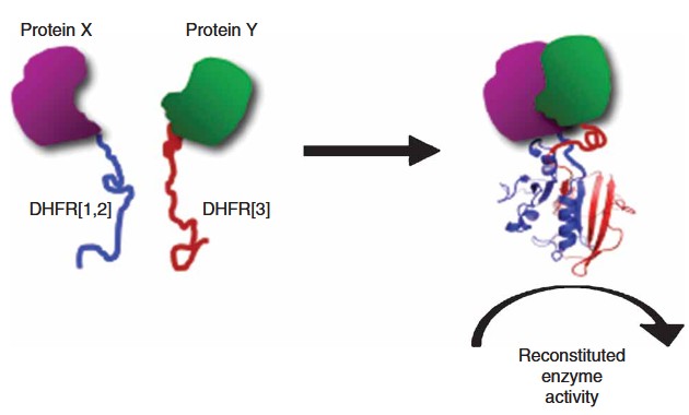 Figure 1. Protein-fragment complementation assay strategy based on the enzyme dihydrofolate reductase. (Remy et al., 2017)