We use cookies to understand how you use our site and to improve the overall user experience. This includes personalizing content and advertising. Read our
Privacy Policy
Transcription factors (TFs) are sequence-specific DNA-binding proteins that regulate gene expression by controlling the transcription of genetic information from DNA to messenger RNA. They are essential for virtually every cellular process, including development, differentiation, immune response, metabolism, and disease pathogenesis. Dysregulation of transcription factor activity has been implicated in cancer, autoimmune disorders, cardiovascular disease, and metabolic syndrome, making them increasingly important as both drug targets and biomarkers.
Understanding where transcription factors bind, how strongly they interact with DNA, and which genes they regulate is fundamental to deciphering gene regulatory networks and identifying therapeutic intervention points. Profacgen offers a comprehensive suite of transcription factor analysis services combining classical biochemical assays with advanced genomic approaches, providing versatile solutions for both prokaryotic and eukaryotic systems.
Background: Transcription Factors as Master Regulators
Transcription factors function through three principal mechanisms that collectively determine gene expression output:
RNA polymerase recruitment and stabilization: TFs bound at promoter-proximal sites directly recruit RNA polymerase II through interactions with the general transcription machinery (TATA-binding protein, TFIID complex) or stabilize the pre-initiation complex, enhancing transcriptional initiation frequency.
Chromatin modification: Many TFs recruit histone-modifying enzymes that alter local chromatin structure. Histone acetyltransferases (HATs) deposit acetyl groups that relax chromatin and promote transcription, while histone deacetylases (HDACs) remove acetyl groups, condensing chromatin and repressing gene expression. This epigenetic layer adds a crucial dimension to transcriptional control.
Coactivator and corepressor recruitment: TFs serve as docking platforms for coactivator complexes (e.g., p300/CBP, mediator) that bridge distal enhancers to the transcription machinery, or corepressor complexes (e.g., NCoR, SMRT) that actively silence transcription. The balance between coactivator and corepressor engagement determines net transcriptional output.
Figure 1. Transcription factors regulate gene expression through RNA polymerase recruitment, chromatin modification, and coactivator/corepressor engagement.
The human genome encodes approximately 1,600–1,800 sequence-specific transcription factors, representing roughly 8% of all protein-coding genes. Among these, an estimated 10% are directly implicated in Mendelian disorders, and many more contribute to complex polygenic diseases. The growing recognition of transcription factors as "undruggable" targets now being drugged—through proteolysis-targeting chimeras (PROTACs), molecular glues, and allosteric inhibitors—has intensified interest in robust TF characterization platforms.
Gene regulatory network mapping: Identify all genomic binding sites for a TF of interest and infer its regulatory network using ChIP-seq combined with RNA-seq
Drug target validation: Confirm that a candidate drug modulates the intended TF-DNA interaction using EMSA or reporter assays
Compound screening: Screen small-molecule libraries for TF modulators using luciferase reporter or EMSA-based formats
Biomarker discovery: Identify TF binding signatures that correlate with disease state, drug response, or patient prognosis
Mechanism-of-action studies: Determine how a compound, genetic perturbation, or environmental stimulus alters TF binding and downstream gene expression
Regulatory element characterization: Map enhancers, silencers, and insulator elements that control tissue-specific or stimulus-responsive gene expression
Why Choose Profacgen?
Comprehensive Assay Portfolio: EMSA, DNase I footprinting, ChIP-qPCR, ChIP-seq, and reporter assays on a single integrated platform.
Genome-Scale Capabilities: ChIP-seq delivers single-nucleotide resolution binding maps across the entire genome.
Quantitative Binding Analysis: EMSA with fluorescence detection provides precise KD measurements for TF-DNA interactions.
Living Cell Context: ChIP and reporter assays preserve native chromatin structure and cellular signaling, capturing physiologically relevant binding.
Bioinformatic Integration: Binding site data are integrated with motif analysis, pathway enrichment, and gene expression correlation to generate actionable biological insights.
Representative Case Studies
Case 1: Genome-Wide Mapping of p53 Binding in Drug-Resistant Lung Cancer
Background:
An oncology research group sought to understand why a subset of lung adenocarcinoma patients developed resistance to MDM2 inhibitors (which activate p53) within 6 months of treatment. They hypothesized that p53 might adopt alternative binding patterns in resistant cells, rewiring its transcriptional program.
Our Solution:
Profacgen performed ChIP-seq for p53 in paired sensitive and resistant cell lines, combined with RNA-seq to correlate binding with gene expression changes. Peak calling, motif analysis, and differential binding analysis were performed using established bioinformatic pipelines. ChIP-qPCR validated 12 candidate differential binding sites.
Final Results:
p53 exhibited a dramatic redistribution in resistant cells: 40% of binding sites gained and 35% lost compared to sensitive cells. The newly acquired sites were enriched near genes involved in epithelial-mesenchymal transition (EMT) and stemness maintenance, while canonical pro-apoptotic targets (BAX, PUMA) showed diminished p53 occupancy. These findings explained the therapeutic failure and suggested combination strategies targeting EMT pathways to restore sensitivity to MDM2 inhibitors.
Case 2: Characterization of a Plant Transcription Factor for Agricultural Biotechnology
Background:
An agricultural biotechnology company had identified a transcription factor (OsDREB1A) that enhanced drought tolerance in rice but did not understand which genes it regulated or how to optimize its activity for commercial varieties.
Our Solution:
Profacgen performed EMSA to confirm DNA binding and determine the optimal binding sequence (RTCGCC), followed by DNase I footprinting to map protected regions within the known dehydration-responsive element (DRE). ChIP-seq in rice seedlings under drought stress identified 847 high-confidence genomic binding sites, and RNA-seq revealed 312 differentially expressed genes associated with OsDREB1A binding.
Final Results:
The integrated analysis defined the OsDREB1A regulon, identifying a core set of 47 genes consistently upregulated by drought-responsive binding. Notably, 12 of these genes encoded previously uncharacterized proteins predicted to function in osmoprotectant synthesis. Overexpression of one such gene (OsOPR3) in a commercial rice variety conferred a 25% yield improvement under controlled drought conditions, providing a direct path to product development.
Q: What is the difference between EMSA and ChIP for studying transcription factor binding?
A: EMSA measures in vitro binding of purified or semi-purified transcription factors to defined DNA probes under controlled conditions. It provides precise quantitative affinity data (KD) and is ideal for comparing binding strengths of different DNA sequences or determining the effects of mutations and compounds on binding. ChIP measures in vivo binding within the native chromatin context of living cells, revealing which genomic sites are actually occupied under physiological conditions. ChIP captures the influence of chromatin accessibility, cooperative binding, and post-translational modifications that EMSA cannot detect. The two methods are highly complementary and are often used together for comprehensive TF characterization.
Q: How much sample do I need for ChIP-seq?
A: For standard ChIP-seq, we typically require 1–5 × 10⁶ cells or 25–50 mg of tissue per immunoprecipitation. For low-input applications (rare cell populations, clinical biopsies), we offer optimized micro-ChIP protocols that achieve robust results from as few as 10,000 cells. During project consultation, we assess your sample availability and recommend the appropriate protocol.
Q: Can you profile transcription factors that lack available antibodies?
A: Yes. For ChIP studies, we can generate epitope-tagged versions of your transcription factor (FLAG, HA, Myc, V5) and use well-validated commercial antibodies against the tag. Alternatively, we can develop custom antibodies specific to your TF. For EMSA, no antibody is required since binding is detected directly by mobility shift of the labeled DNA probe.
Q: What bioinformatic analysis is included with ChIP-seq?
A: Our standard ChIP-seq analysis pipeline includes: peak calling with MACS2 or equivalent, peak annotation to nearest genes and genomic features, de novo and known motif enrichment analysis, differential binding analysis (if multiple conditions), GO and pathway enrichment for target genes, and integration with RNA-seq data (if available). All results are delivered as publication-ready figures and comprehensive data tables.
Q: Can you screen compound libraries for transcription factor modulators?
A: Yes. We have developed high-throughput screening formats based on luciferase reporter assays and AlphaScreen-based EMSA for screening small-molecule libraries against transcription factor-DNA interactions. These formats are compatible with 384- and 1536-well plates and can screen 10,000–100,000 compounds per campaign. Confirmed hits are validated by orthogonal assays (EMSA, SPR, ITC) to eliminate false positives.
Online Inquiry
Fill out this form and one of our experts will respond to you within one business day.

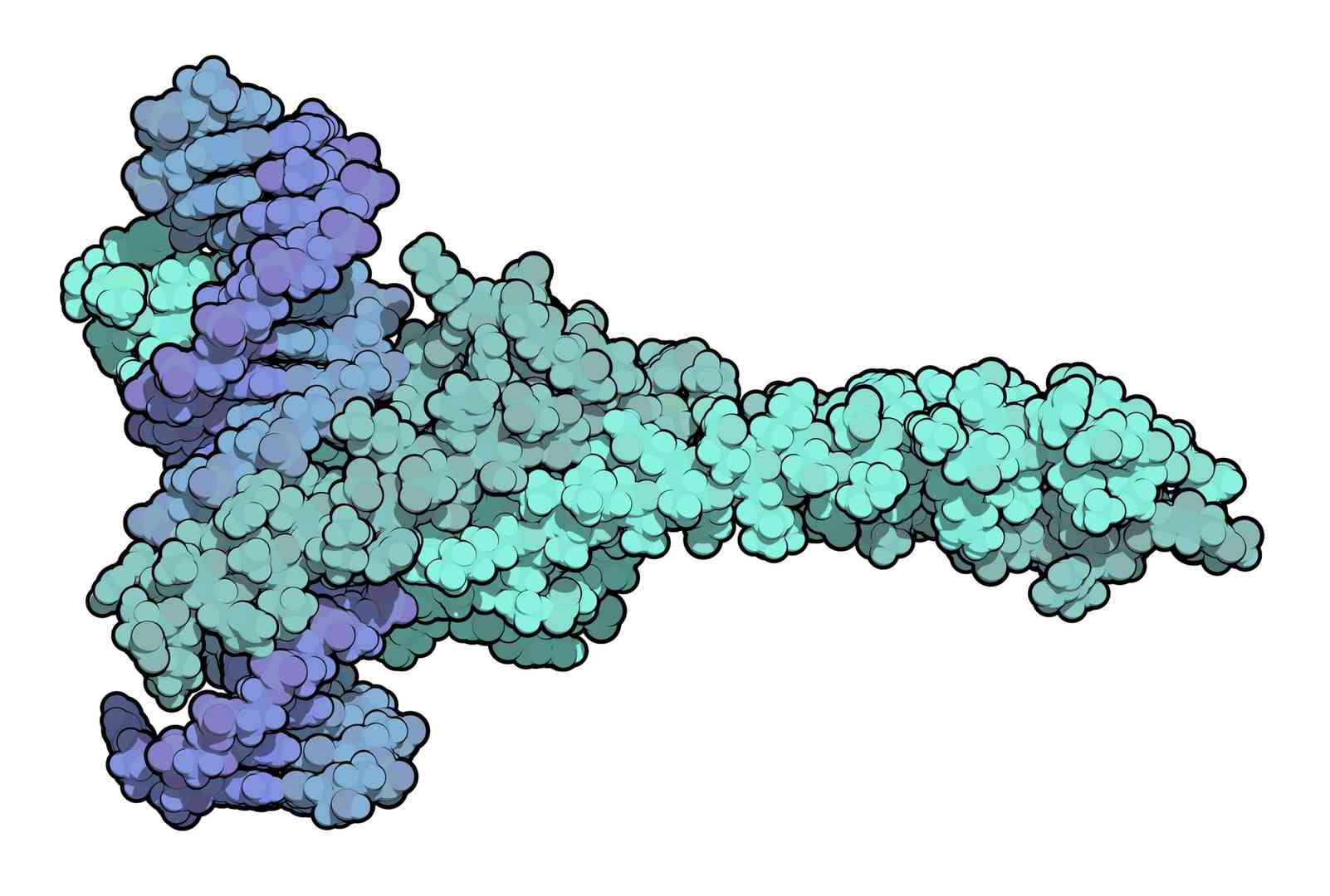
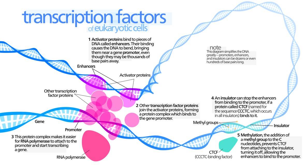 Figure 1. Transcription factors regulate gene expression through RNA polymerase recruitment, chromatin modification, and coactivator/corepressor engagement.