The complement system is an ancient and evolutionarily conserved element of innate immunity, comprising over 50 plasma and membrane-associated proteins that orchestrate host defense against bacterial, viral, and fungal pathogens. These proteins circulate as inactive precursors and undergo sequential proteolytic activation through three distinct pathways—classical, alternative, and lectin—ultimately converging on the formation of the Membrane Attack Complex (MAC) and generation of potent pro-inflammatory anaphylatoxins. Because the complement system operates as a highly effective killing mechanism, its activity is tightly controlled by soluble regulators and membrane-bound inhibitory receptors. Disruption of this regulatory balance, whether through excessive, uncontrolled, or deficient activation, drives the pathogenesis of autoimmune disorders, inflammatory diseases, and thrombotic microangiopathies. Emerging evidence also implicates complement in tumor immune evasion and progression, underscoring the importance of precise complement analysis in drug development, clinical research, and immunotherapy. Profacgen provides a comprehensive Complement Assays platform that integrates component quantification, functional pathway evaluation, and cytotoxicity assessment to support therapeutic discovery, biomarker validation, and regulatory compliance.
The complement cascade serves as a critical bridge between innate recognition and adaptive immune responses. Upon activation, proteolytic cleavage of precursor proteins generates enzymatic complexes that amplify inflammatory signals, opsonize microbial surfaces for phagocytic clearance, and directly lyse susceptible targets through pore-forming MAC insertion.
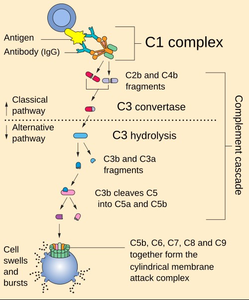
Each activation pathway is initiated by distinct molecular recognition events: the classical pathway by antibody-antigen complexes or C-reactive protein binding to C1q; the lectin pathway by mannose-binding lectin (MBL) or ficolins recognizing carbohydrate patterns on microbial surfaces; and the alternative pathway by spontaneous C3 hydrolysis (tick-over) and Factor B/D-mediated amplification on activating surfaces. Despite divergent initiation mechanisms, all three pathways generate homologous C3 convertase enzymes that cleave C3 into C3a and C3b, with C3b subsequently participating in C5 convertase assembly to drive C5 cleavage and terminal pathway activation.
The biological consequences of complement activation extend beyond direct pathogen elimination. C3a and C5a function as potent chemoattractants recruiting neutrophils, monocytes, and mast cells to sites of inflammation. C3b and its degradation products iC3b and C3dg serve as opsonins recognized by complement receptors on phagocytes, facilitating microbial engulfment and antigen presentation. Meanwhile, MAC formation disrupts membrane integrity, causing osmotic lysis of Gram-negative bacteria and susceptible host cells.
Given the destructive potential of unregulated complement, multiple control mechanisms operate at each cascade step. C1 inhibitor (C1-INH) dissociates C1r and C1s from C1q; Factor H and Factor I collaboratively degrade C3b; CD55 (decay-accelerating factor) and CD59 (protectin) protect host cells from autologous complement attack. When these regulatory mechanisms fail, complement-mediated tissue injury ensues—exemplified by paroxysmal nocturnal hemoglobinuria (PNH, CD55/CD59 deficiency), atypical hemolytic uremic syndrome (aHUS, Factor H dysfunction), and hereditary angioedema (C1-INH deficiency).
Profacgen has established a fully integrated complement analysis platform that combines quantitative immunoassays, functional pathway activity measurements, and cell-based cytotoxicity evaluation. Our services are performed in compliance with GLP and ISO 17025 standards, ensuring data integrity and regulatory readiness for preclinical and clinical applications.
Our complement assays team brings deep expertise in complement biochemistry and therapeutic development. We provide personalized assay design, execution, and data interpretation tailored to each project's scientific and regulatory requirements. Whether profiling complement activation in autoimmune disease models, validating potency assays for complement-targeted therapeutics, or monitoring immunogenicity in biologic development programs, we deliver high-quality analytical results with comprehensive documentation.
Complement Activation Assays
Our activation assays detect pathway-specific engagement and terminal complex formation using neoantigen-specific antibodies that recognize cleavage-dependent epitopes absent from native precursor proteins.
Complement Quantification Assays
Our quantification services provide precise measurement of individual complement protein concentrations using validated immunoassays with demonstrated specificity and freedom from cross-reactivity.
Cytotoxicity Assays
Our cell-based cytotoxicity assays measure the functional endpoint of complement-mediated killing, providing direct evidence of MAC formation and membrane disruption.
Profacgen offers two focused complement assay platforms designed to address distinct analytical needs in research and therapeutic development. Each service line provides specialized expertise and validated methodologies tailored to its specific application domain.
Precise quantification of individual complement proteins, cleavage fragments, and regulatory factors using ELISA, nephelometry, and multiplex detection. Ideal for biomarker discovery, deficiency diagnosis, therapeutic target engagement assessment, and pathway consumption profiling.
Learn more about Complement Component Assays →
Biological activity assessment of classical, alternative, and MB-lectin pathways through hemolytic and solid-phase functional readouts. Essential for therapeutic potency testing, clinical complement function monitoring, off-target reactivity evaluation, and inherited deficiency characterization.
Learn more about Functional Complement Assays →
Antibody Therapeutics Development
Complement assays are integral to the development and characterization of therapeutic monoclonal antibodies that leverage or suppress complement effector functions. We support:
Immunogenicity Assessment
Unwanted complement activation represents a critical safety concern for biologic drugs, cell therapies, and nanomedicine formulations. Our immunogenicity services include:
Inflammatory Disease Research
Complement dysregulation is a hallmark of numerous autoimmune and inflammatory conditions. Our assays enable:
Background:
A biosimilar developer required comprehensive complement effector function characterization for an anti-CD20 candidate referencing a marketed originator product. Regulatory guidelines demanded demonstration of comparable C1q binding, C3b deposition, and CDC activity to establish biosimilarity. Initial in-house assays showed inconsistent CDC readouts across donor serum lots, threatening program timelines.
Our Solution:
Profacgen implemented a three-tier complement characterization package: (1) C1q binding affinity by SPR; (2) C3b deposition on CD20-positive target cells using a neoantigen-specific detection antibody; and (3) standardized CDC potency assays with qualified human serum pools as complement source. We developed a normalized CDC assay format controlling for inter-donor complement variability, enabling robust statistical comparison between biosimilar and originator.
Final Results:
The integrated data package demonstrated equivalent complement-mediated effector function within predefined biosimilarity margins (90% CI within 80–125%). The C3b deposition and CDC potency assays were qualified for QC lot release testing, supporting the client's BLA submission and successful regulatory approval.
Background:
An academic research group investigating SLE pathogenesis hypothesized that lectin pathway dysregulation—specifically MBL variant-driven excessive activation—contributed to disease flare patterns and organ damage accrual independent of classical pathway consumption. Existing clinical assays measured only total C3 and C4, providing insufficient granularity to test this hypothesis.
Our Solution:
Profacgen designed a prospective biomarker study employing parallel functional and quantitative assays: MBL pathway-specific functional activity (mannan-coated activation with C5b-9 detection), MBL concentration and structural variant quantification, and multiplex profiling of C2, C4b, C5a, Factor I, and MASP-2. Samples from 200 SLE patients and 100 healthy controls were analyzed in a blinded, batch-randomized format with rigorous QC monitoring.
Final Results:
The study identified a distinct SLE patient subset (18% of cohort) with elevated lectin pathway functional activity despite normal total C4, associated with specific MBL2 gene variants and higher Systemic Lupus Erythematosus Disease Activity Index (SLEDAI) scores. This biomarker signature was subsequently validated in an independent cohort and published in a high-impact rheumatology journal, advancing the field toward pathway-specific SLE stratification and targeted MASP inhibitor development.
References:
Fill out this form and one of our experts will respond to you within one business day.