
Protein microarray technology enables high-throughput, parallel screening of thousands of protein interactions on a miniature solid surface. Profacgen provides comprehensive protein microarray services for detecting protein-protein, protein-DNA, protein-RNA, and protein-small molecule interactions. Our platform supports direct detection, sandwich assay, reverse-phase array, and competition-based formats, delivering quantitative binding data with minimal sample consumption.
A protein microarray consists of thousands of distinct proteins or protein variants spotted in an ordered pattern onto a chemically modified glass slide or membrane substrate. The array is incubated with a fluorescently labeled or tagged query sample (purified protein, cell lysate, serum, or small molecule), and binding events are detected by fluorescence scanning. The spatial position of each spot encodes the identity of the immobilized protein, enabling rapid identification of interaction partners across the entire proteome represented on the chip.
Multiple detection configurations expand the range of detectable interactions:
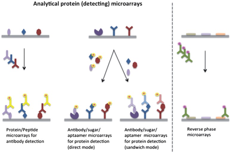 Figure 1. Protein microarray technology. (Cretich et al., 2014)
Protein microarrays achieve throughput unmatched by pairwise methods, screening thousands of interactions in a single experiment with microgram-scale protein consumption.
Profacgen provides comprehensive protein microarray screening services for research and diagnostic applications. Our offerings include:
| Service Component | Description |
|---|---|
| Protein-Protein Interaction Screening | Probe protein microarrays with your purified or labeled protein of interest. Direct fluorescence detection; up to 16,000 proteins per slide. Deliverables: binding profile, hit list with quantified fluorescence data, full report. |
| Serum Autoantibody Profiling | Screen patient sera against proteome microarrays to identify disease-associated autoantibodies. Dual-color detection with statistical analysis. Deliverables: autoantibody profile, differential analysis, biomarker panel. |
| Small Molecule Binding Screen | Identify protein targets of small molecules by competition or direct binding assays. Target deconvolution for phenotypic screening hits. Deliverables: target protein hits, binding affinity estimates, selectivity analysis. |
| Custom Microarray Fabrication | Print your protein set or our curated collections onto standard or specialized substrates. Multiple surface chemistries available. Deliverables: printed microarray, QC validation, titer assessment. |

To complement your protein interaction analysis, explore our comprehensive portfolio of related screening and profiling services.
Background:
A research institute studying SARS-CoV-2 pathogenesis needed to comprehensively identify human proteins interacting with the viral nucleocapsid (N) protein, a key structural component involved in viral RNA packaging and replication. Understanding the full host-virus interaction network was critical for elucidating viral replication mechanisms, identifying potential therapeutic targets, and uncovering host factors that could be exploited for antiviral strategies.
Our Solution:
Using our human proteome microarray containing over 20,000 full-length proteins expressed in their native conformations, we probed the array with fluorescently labeled recombinant N protein at systematically optimized concentrations to ensure specific binding while minimizing nonspecific background. Binding events were detected by dual-color fluorescence scanning, enabling precise quantification of signal intensities. Stringent background subtraction and robust statistical filtering—including Z-score normalization and false discovery rate (FDR) control—were applied to distinguish true interactors from technical noise and nonspecific binders.
Final Results:
The screen identified 341 specific interactors, including 12 previously uncharacterized binders. Gene ontology analysis revealed enrichment in RNA processing and stress granule pathways. The top 20 hits were independently validated by co-immunoprecipitation with a validation rate of 85%, and three interactions were subsequently confirmed as essential for viral RNA packaging.
Background:
A clinical diagnostics company sought to identify novel autoantibody signatures for early detection of rheumatoid arthritis (RA), an autoimmune disorder where timely intervention significantly improves patient outcomes. Existing clinical biomarkers—including RF and anti-CCP—lacked sufficient sensitivity for pre-symptomatic screening and failed to capture the full heterogeneity of RA patient autoantibody repertoires. A multiplexed, unbiased proteome-wide approach was urgently needed to discover new biomarker candidates capable of detecting RA at its earliest, most treatable stages.
Our Solution:
We screened sera from 120 RA patients and 120 matched healthy controls on our human proteome microarray platform. Each serum sample was incubated on separate arrays, and dual-color detection using Cy3-labeled anti-human IgG enabled quantitative, side-by-side comparison of autoantibody binding levels across all 20,000+ immobilized proteins simultaneously. Differential signal analysis with robust statistical methods—including moderated t-tests and multiple hypothesis correction—was employed to identify targets with significant abundance changes between patient and control groups.
Final Results:
Comparative analysis identified a panel of 47 autoantibody targets significantly elevated in RA patients, including 15 not previously associated with the disease. A classifier combining 8 top markers achieved 91% sensitivity and 87% specificity in a blinded validation cohort. The client is progressing the marker panel toward clinical assay development.
Get a Quote for your microarray project
References:
Fill out this form and one of our experts will respond to you within one business day.