
SILAC (Stable Isotope Labeling by Amino Acids in Cell Culture) combined with immunoprecipitation and mass spectrometry (SILAC-IP-MS) represents the gold standard for quantitative analysis of protein-protein interactions in living cells. By metabolically incorporating heavy isotope-labeled amino acids into proteins during normal cell growth, SILAC enables highly accurate, reproducible quantification of protein abundance ratios between experimental and control conditions directly at the MS level—eliminating the variability associated with chemical labeling or label-free approaches and providing unmatched statistical power for distinguishing specific interactors from background contaminants.
When applied to immunoprecipitation, SILAC provides an internal standard in every sample: cells expressing the bait protein are grown in heavy medium (containing 13C6-L-lysine and 13C6,15N4-L-arginine), while control cells expressing an irrelevant protein or empty vector are grown in light medium (natural isotopes). After mixing equal protein amounts from heavy and light cultures and performing immunoprecipitation, specific interactors show a high heavy-to-light (H/L) ratio because they are enriched with the bait, whereas non-specific contaminants show a ratio near 1 because they bind equally to both samples. This quantitative discrimination is both extraordinarily sensitive and remarkably robust. Profacgen has established a streamlined SILAC-IP-MS platform that delivers publication-quality interaction data with rigorous statistical validation.
Stable Isotope Labeling by Amino Acids in Cell Culture combined with Immunoprecipitation and Mass Spectrometry (SILAC-IP-MS) is a quantitative proteomic approach for precisely distinguishing true protein–protein interactors from nonspecific background binders. By incorporating stable isotope-labeled amino acids (e.g., 13C6-arginine and 13C6-lysine) into the proteome of cultured cells during metabolic labeling, SILAC enables accurate relative quantitation between experimental and control samples within a single mass spectrometric run.
In a typical SILAC-IP-MS workflow, "heavy"-labeled cells expressing the bait protein are compared against "light"-labeled control cells (or vice versa). Both populations are lysed, subjected to immunoprecipitation using an antibody targeting the bait or its epitope tag, and the captured protein complexes are pooled prior to LC-MS/MS analysis. True interactors—proteins that associate specifically with the bait—exhibit a heavy-to-light (H/L) ratio significantly greater than 1 (or less than 1 in a reverse labeling scheme), while background contaminants that bind nonspecifically to the antibody or resin display a ratio near 1. This built-in quantitative filter effectively eliminates false positives arising from common IP artifacts, providing high-confidence interactor identification without the need for extensive empirical optimization of wash stringency.
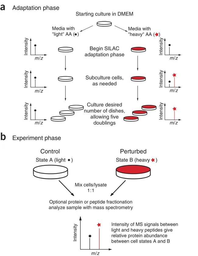 Figure 1. Overview of SILAC protocol.The SILAC experiment consists of two distinct phases—an adaptation (a) and an experimental (b) phase. (Ong amd Mann, 2006)
SILAC-IP-MS is particularly valuable for studying dynamic interactions—such as those regulated by post-translational modifications, signaling stimuli, or drug treatment—as well as for comparing interactome changes across different cellular states. The method is applicable to any cell line capable of efficient metabolic labeling and is compatible with most IP-compatible tags and antibodies. Profacgen's SILAC-IP-MS platform delivers quantitatively rigorous, publication-ready interactome data with minimal background interference.
Exceptional Quantitation Accuracy
Metabolic labeling achieves coefficient of variation (CV) typically below 10% across replicates—2–3-fold better than chemical labeling or label-free methods. This precision enables detection of even subtle interaction changes.
Internal Standard in Every Sample
Because heavy and light cells are mixed before any sample handling, quantitative ratios are unaffected by variability in IP efficiency, sample loss, or MS instrument performance—providing unmatched reproducibility.
Complete Labeling Efficiency
After 6–8 cell doublings in SILAC medium, >99% of proteins incorporate the labeled amino acids, eliminating the partial-labeling artifacts that complicate chemical labeling approaches.
Statistical Rigor
Quantitative H/L ratios enable objective, statistics-based filtering of interactors using established algorithms (SAINT, CompPASS), replacing subjective band-intensity judgments with reproducible, data-driven decisions.
| Step | Description | Timeline |
|---|---|---|
| 1. SILAC Adaptation | Cells are cultured in SILAC medium (heavy: 13C6-lysine, 13C615N4-arginine; light: natural isotopes) for ≥6 doublings to achieve >98% labeling efficiency. Labeling is verified by MS. | 2–3 weeks |
| 2. Bait Expression | Bait protein (epitope-tagged or endogenous with specific antibody) is expressed in heavy cells. Control cells (light) express empty vector or unrelated bait. | 1 week |
| 3. Cell Lysis & IP | Equal protein amounts from heavy and light cultures are combined and subjected to immunoprecipitation under conditions preserving native complexes. | 2–3 days |
| 4. On-Bead Digestion & MS | Captured proteins are digested with trypsin directly on beads. Peptides are analyzed by nano-LC-MS/MS on an Orbitrap instrument with high-resolution acquisition. | 1–2 weeks |
| 5. Data Analysis | MaxQuant quantifies H/L ratios for every identified protein. SAINTexpress scores specificity. GO enrichment and network analysis provide biological context. | 2–3 weeks |
Forward SILAC (Standard)
Bait in heavy, control in light.
Reverse SILAC
Bait in light, control in heavy.
Triple SILAC
Light, medium, and heavy labels in one experiment.
To complement your protein interaction analysis, explore our comprehensive portfolio of related screening and profiling services.
Background:
An oncology team studying vemurafenib resistance in BRAFV600E melanoma observed that resistant cells maintained MAPK pathway activity despite BRAF inhibition. They hypothesized that BRAF had acquired novel protein interactions that bypassed the drug block.
Our Solution:
Profacgen performed triple SILAC-IP-MS of FLAG-BRAFV600E: sensitive cells (light), resistant cells (medium), and empty vector control (heavy). BRAF was immunoprecipitated from each culture, mixed at 1:1:1 ratio, and analyzed by LC-MS/MS. MaxQuant quantified L/H and M/H ratios; SAINTexpress scored specificity.
Final Results:
Triple SILAC identified 15 proteins with >3-fold increased association with BRAF in resistant cells. The most significant was CRAF (MAP3K3), which showed a 6-fold increase—indicating BRAF-CRAF heterodimerization as a resistance mechanism. Notably, EGFR was identified as a novel BRAF interactor only in resistant cells, suggesting RTK-mediated reactivation. Combined BRAF + EGFR inhibition with vemurafenib + erlotinib restored sensitivity in resistant cells and achieved a 78% tumor reduction in a patient-derived xenograft model. The data supported a Phase I combination trial.
Background:
A neurodevelopmental genetics group had identified de novo mutations in SHANK3—a postsynaptic scaffold protein—in patients with autism spectrum disorder (ASD). They needed to determine how these mutations altered SHANK3's synaptic protein interaction network.
Our Solution:
Profacgen performed forward-reverse SILAC-IP-MS of wild-type and R12C mutant SHANK3. Forward: WT-SHANK3 (heavy) vs. empty vector (light). Reverse: R12C-SHANK3 (heavy) vs. empty vector (light). GFP-Trap magnetic beads captured GFP-tagged SHANK3 from HEK293 cells. Combined analysis identified WT-specific, mutant-specific, and shared interactors.
Final Results:
Forward SILAC identified 62 WT-SHANK3 interactors (SAINT >0.95), including 18 known PSD proteins. Reverse SILAC identified 47 R12C-SHANK3 interactors, with 14 proteins showing >3-fold differential binding compared to WT. The R12C mutation selectively disrupted interactions with HOMER1 and SHANK1 (cytoskeletal organizers) while preserving binding to NMDA receptor subunits. This selective disruption impaired dendritic spine maturation in iPSC-derived neurons. The data provided a mechanistic link between SHANK3 mutations and altered synapse development in ASD.
Start Your SILAC-IP-MS Project
References:
Fill out this form and one of our experts will respond to you within one business day.