We use cookies to understand how you use our site and to improve the overall user experience. This includes personalizing content and advertising. Read our
Privacy Policy
Reversible protein phosphorylation is the most widespread and functionally critical post-translational modification in eukaryotic cells. Radioisotope-labeling studies have demonstrated that approximately 30% of all cellular proteins can be phosphorylated, and this dynamic modification regulates virtually every aspect of cell physiology—from energy metabolism and protein synthesis to gene expression, cytoskeletal remodeling, muscle contraction, and the release of signaling factors. Dysregulated phosphorylation is a hallmark of cancer, inflammatory diseases, and metabolic disorders, making phosphoprotein profiling an indispensable tool for drug discovery, target validation, and mechanism-of-action studies.
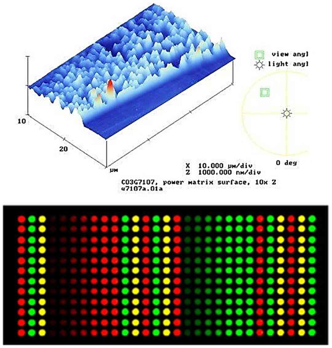
Profacgen offers comprehensive signal pathway phosphorylation profiling using high-density phosphorylated antibody microarrays. Our proprietary platform integrates 1,318 highly specific antibodies targeting 679 phosphorylation sites on 432 signaling proteins, enabling simultaneous monitoring of activation states across multiple pathways in a single experiment. Each phosphorylation site is interrogated by a matched pair of antibodies recognizing the phosphorylated and non-phosphorylated forms, ensuring sensitive, specific, and quantitative detection of signaling dynamics in tissue, cell, and fluid samples.
Background: Protein Phosphorylation and Signal Transduction
Protein phosphorylation was first described in the 1950s by Edmond Fischer and Edwin Krebs, who discovered that the conversion of glycogen phosphorylase b to the active a form was catalyzed by a kinase and reversed by a phosphatase. This groundbreaking work earned them the Nobel Prize in Physiology or Medicine in 1992 and established phosphorylation as a fundamental regulatory mechanism. Today, the human kinome comprises approximately 538 protein kinases and nearly 160 protein phosphatases, which together constitute one of the largest and most therapeutically relevant enzyme families in the genome.
Phosphorylation functions as a molecular switch, rapidly altering protein activity, subcellular localization, protein-protein interactions, and stability in response to extracellular cues. A single growth factor binding to its receptor can trigger a phosphorylation cascade involving hundreds of proteins within minutes, transmitting signals from the plasma membrane to the nucleus with remarkable speed and specificity. This network architecture underlies cellular processes as diverse as proliferation, survival, differentiation, migration, and apoptosis.
The advent of phospho-specific antibodies in the 1990s and protein microarray technology in the early 2000s transformed phosphoprotein analysis from labor-intensive, single-target Western blots to high-throughput, multiplexed profiling. Protein microarrays enable the parallel interrogation of hundreds of phosphorylation sites using minimal sample input, dramatically accelerating the pace of signal transduction research. Profacgen's phosphorylated antibody chip platform builds on these technological foundations, offering researchers a powerful tool for comprehensive pathway analysis in drug discovery and translational research.
Platform Specifications and Technical Features
Profacgen's phosphorylated antibody microarrays are manufactured under stringent quality-controlled conditions to ensure reproducibility, sensitivity, and specificity:
Parameter
Specification
Substrate format
1 × 25 × 76 mm glass slide with three-dimensional polymer membrane coating
Spot diameter
~60 μm
Spotting volume
0.5–1 nL per feature
Antibody content
1,318 antibodies (paired phospho- and total-antibodies) targeting 679 phosphorylation sites on 432 signaling proteins
Pathway coverage
AKT, CREB, Cell Cycle, Cytoskeletal Signaling, GPCR, JAK/STAT, MAPK/ERK, NF-κB, PI3K, TGF-β, Wnt/β-catenin, and more
Technical replicates
6 replicates per antibody spot
Detection method
Fluorescence (single- or multi-color labeling); compatible with Cy3, Cy5, Alexa Fluor dyes
All capture antibodies expressed and purified from a eukaryotic yeast system under native, non-denaturing conditions
Production environment
Temperature 4–8 °C, humidity 30–40%; samples fixed overnight at 4 °C; long-term storage at −80 °C
Signal Pathway Panels
Our phosphorylated antibody chip platform provides comprehensive coverage of the major signaling pathways implicated in human disease and therapeutic intervention:
AKT Pathway Phosphorylation Chip
Monitor the PI3K/AKT/mTOR axis—the most frequently activated survival pathway in cancer.
Kinase inhibitor mechanism-of-action studies: Determine which signaling pathways are selectively inhibited or activated by your compound, revealing on-target and off-target effects at the pathway level
Drug resistance profiling: Identify compensatory phosphorylation events that emerge upon chronic drug exposure, informing rational combination strategies to overcome resistance
Biomarker discovery for patient stratification: Identify baseline phosphoprotein signatures that predict therapeutic response or resistance, supporting precision medicine approaches
Oncology drug discovery: Profile pathway activation across cancer cell lines and patient-derived xenograft models to identify driver pathways and therapeutic vulnerabilities
Immuno-oncology research: Monitor immune checkpoint signaling, T-cell receptor proximal signaling, and cytokine-driven JAK/STAT activation in response to immunotherapeutic agents
Compound safety assessment: Detect off-target kinase inhibition and unintended pathway activation that may predict adverse events in clinical development
Disease mechanism elucidation: Compare phosphoprotein landscapes between diseased and healthy tissues to identify novel drug targets and diagnostic markers
Why Choose Profacgen?
Unmatched Coverage: 1,318 antibodies targeting 679 phosphorylation sites on 432 proteins—comprehensive pathway profiling in a single experiment.
Phospho-Total Pairs: Matched antibodies for phosphorylated and non-phosphorylated states enable precise quantification of activation ratios, not just presence.
High Reproducibility: Six technical replicates per spot, stringent production QC, and validated yeast-expressed antibodies ensure consistent, reliable data.
Minimal Sample Input: Comprehensive profiling from as little as 50–100 μg of protein lysate, preserving precious clinical specimens.
End-to-End Support: From experimental design and sample processing to data analysis and pathway interpretation, our scientists guide you at every step.
Integrated Analysis: Raw data are processed, normalized, and interpreted with pathway enrichment analysis and heat map visualization included with every project.
Representative Case Studies
Case 1: Multi-Pathway Rewiring in BRAF-Mutant Melanoma upon MEK Inhibitor Treatment
Background:
A pharmaceutical company developing a next-generation MEK1/2 inhibitor for BRAFV600E melanoma observed that a subset of patient-derived cell lines developed resistance within 4 weeks of chronic treatment. They needed to identify the compensatory signaling events driving resistance to inform combination therapy design.
Our Solution:
Profacgen performed full-panel phosphorylated antibody chip profiling on 6 paired parental and resistant cell lines at baseline and 1, 7, 14, and 28 days after MEK inhibitor exposure. Differential phosphorylation analysis was performed using limma with empirical Bayes moderation, and pathway enrichment was assessed using ReactomePA.
Final Results:
Time-course analysis revealed progressive upregulation of PI3K/AKT signaling beginning at day 7, with AKT S473 phosphorylation increasing 5-fold by day 28. Concomitant activation of FGFR1 and IGF1R autocrine loops was detected. The combination of MEK + PI3K inhibitors restored sensitivity in all 6 resistant lines, with synergistic effects confirmed by Bliss independence analysis. These data directly informed the design of a Phase Ib combination trial (NCT037_XXXXX).
Case 2: Baseline Phosphoprotein Signature Predicts Response to EGFR Inhibition in NSCLC
Background:
An academic medical center running a Phase II trial of a third-generation EGFR tyrosine kinase inhibitor (TKI) in EGFR-mutant non-small cell lung cancer (NSCLC) sought a predictive biomarker to identify patients likely to achieve durable responses (>12 months).
Our Solution:
Profacgen performed baseline phosphorylated antibody chip profiling on pre-treatment tumor biopsies from 68 enrolled patients. Machine learning (LASSO logistic regression with 10-fold cross-validation) was applied to identify a minimal phosphoprotein signature predictive of durable response (PFS >12 months vs. ≤6 months).
Final Results:
A 5-phosphoprotein signature (low p-STAT3 Y705, high p-ERK T202/Y204, low p-AKT S473, high p-EGFR Y1068, low p-Src Y416) predicted durable response with 89% sensitivity and 82% specificity (AUC = 0.91). Patients classified as "high-responder" by the signature achieved a median PFS of 18.4 months vs. 4.2 months in "low-responder" patients (HR = 0.18, p < 0.001). The signature is being prospectively validated in an ongoing Phase III trial.
Q: How does phosphorylated antibody chip compare to mass spectrometry-based phosphoproteomics?
A: Phosphorylated antibody chips offer several advantages over MS-based phosphoproteomics: (1) Superior sensitivity for low-abundance phosphoproteins due to antibody enrichment; (2) No enrichment bias toward highly phosphorylated or abundant proteins; (3) Faster turnaround (days vs. weeks) due to streamlined sample processing; (4) Quantitative phospho/total ratios from matched antibody pairs; and (5) Lower cost per sample for targeted pathway panels. MS phosphoproteomics offers the advantage of discovering novel phosphorylation sites. The two approaches are highly complementary: antibody chips excel for targeted, quantitative pathway monitoring, while MS is preferred for unbiased discovery of new phosphosites.
Q: What sample types and amounts are required?
A: Our phosphorylated antibody chips are compatible with a wide range of sample types including cultured cell lysates (50–100 μg protein per chip), fresh-frozen or FFPE tissue lysates (100–200 μg), xenograft and PDX samples, and body fluids (serum, plasma, CSF). For optimal results, we recommend preparing lysates in RIPA or a suitable phosphatase inhibitor-containing buffer, followed by protein quantification (BCA or Bradford). We provide detailed sample preparation protocols upon project initiation.
Q: Can the chip detect phosphorylation changes in clinical biopsy samples?
A: Yes. The platform has been successfully applied to clinical tumor biopsies, core needle biopsies, and fine-needle aspirate samples. For FFPE tissues, we have optimized antigen retrieval and de-crosslinking protocols that preserve phospho-epitopes. Sensitivity is typically sufficient to detect 2-fold changes in phosphorylation with >90% statistical power when starting with 100–200 μg of total protein. We recommend including biological replicates (n ≥ 3 per group) to ensure statistical robustness.
Q: How is the data analyzed and what deliverables are provided?
A: Every project includes a complete data analysis package: (1) raw and background-subtracted fluorescence intensity values for all 1,318 spots; (2) phospho/total signal ratios normalized to internal controls; (3) differential phosphorylation analysis (fold-change, p-values, FDR-corrected q-values); (4) hierarchical clustering and heat map visualization; (5) pathway enrichment analysis (KEGG, Reactome); and (6) a comprehensive report with methods, results, and biological interpretation. All raw data files are provided in standard formats (CSV, Excel) for further analysis.
Q: Can I request a custom phosphorylation panel focused on specific pathways?
A: Yes. In addition to our standard full-panel chip, we offer custom sub-panels focused on specific pathways of interest (e.g., PI3K/AKT only, MAPK only, or a combination of 2–3 pathways). Custom panels reduce cost and analysis time when comprehensive profiling is not required. We can also incorporate additional phospho-specific antibodies not present on the standard chip, subject to antibody availability and validation. Contact us to discuss your specific pathway monitoring requirements.
Q: How long does a typical phosphorylation profiling project take?
A: A standard phosphorylated antibody chip project is typically completed in 2–3 weeks from sample receipt: sample QC and processing (2–3 days), chip hybridization and scanning (3–4 days), and data analysis and report generation (5–7 days). Projects involving large sample cohorts (>50 samples) or complex experimental designs (time-course, dose-response) may require 4–6 weeks. Expedited processing is available for time-critical projects.
Q: Are the antibodies on the chip cross-reactive between species?
A: Most antibodies on our phosphorylated antibody chip are raised against human protein sequences and have been validated for cross-reactivity with mouse, rat, and monkey orthologs when the target phospho-epitope is conserved. We provide a species cross-reactivity chart with every chip, indicating which antibodies are expected to perform reliably in non-human samples. For projects using model organisms, we recommend confirming key findings with species-specific orthogonal assays.
References:
Tu S, Jiang HW, Liu CX, Zhou SM, Tao SC. Protein microarrays for studies of drug mechanisms and biomarker discovery in the era of systems biology. Curr Pharm Des. 2014;20(1):49-55. doi:10.2174/138161282001140113123707
Online Inquiry
Fill out this form and one of our experts will respond to you within one business day.