We use cookies to understand how you use our site and to improve the overall user experience. This includes personalizing content and advertising. Read our
Privacy Policy
At Profacgen, our protein biotinylation service provides comprehensive, high-quality biotin labeling of proteins, antibodies, peptides, and other biomolecules for applications spanning from basic research to diagnostic and therapeutic development. Biotinylation—the covalent attachment of biotin (vitamin H) to a target molecule—is one of the most widely used protein modification strategies in the life sciences, leveraging the extraordinarily high affinity of biotin for avidin and streptavidin to enable versatile capture, detection, and targeting applications.
Profacgen has been providing protein biotinylation services for years and has delivered custom biotinylation at a variety of scales for both academic and industrial customers. Our platform offers multiple chemistries for site-selective biotin attachment, including amine-reactive, thiol-reactive, and carboxyl-reactive strategies, each optimized to preserve the biological activity of the target protein while achieving efficient, reproducible labeling. Beyond conjugation, we provide a complete suite of supporting services including immobilization on streptavidin surfaces, purification of biotinylated species, and comprehensive analytical characterization to ensure that every batch meets your exact specifications.
Background: The Biotin-Streptavidin System
The biotin–streptavidin system is one of the most widely used affinity pairs in biotechnology. Streptavidin (52.8 kDa tetramer) harbors four high-affinity biotin-binding pockets, each burying ~800 Å2 of surface area through extensive H-bonding, van der Waals contacts, and water-mediated interactions—conferring exceptional affinity (KD ~10-15) and specificity.
Key advantages include: near-irreversible binding under physiological conditions, enabling capture at sub-picomolar levels; high specificity tolerant of detergents and chaotropes; rapid association (~107 M-1s-1); stability across pH 2–13, organics, and up to 80°C; and broad compatibility with enzymes, fluorophores, magnetic beads, gold nanoparticles, and solid surfaces.
Biotin placement on the protein is critical. Short spacers can sterically hinder streptavidin access, reducing effective affinity 10–100‑fold, whereas flexible linkers (e.g., PEG4, PEG11, LC-linkers) project biotin outward for unobstructed binding. Profacgen offers diverse spacer options to optimize performance for each application.
Figure 1. Biotin–streptavidin system. (Feng and Zhu, 2019)
Our Biotinylation Methods
Profacgen provides three complementary biotinylation chemistries, each suited to different protein contexts and application requirements:
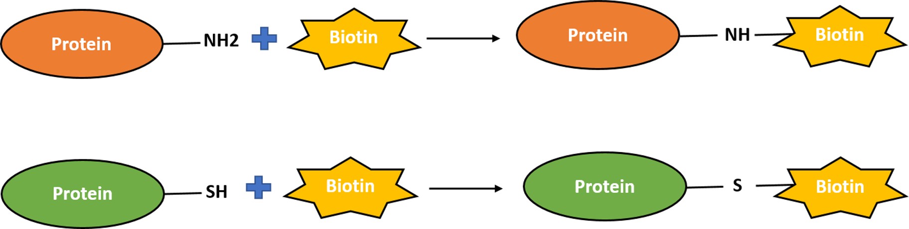
Primary Amine Biotinylation
The most widely used and broadly applicable biotinylation strategy, targeting the ε-amino groups of lysine residues and the α-amino group at the N-terminus.
Reaction at pH 4.5–6.0 (MES buffer); 1–2 hours at RT
Complementary to amine-targeted strategies; useful for acidic proteins
Best for: proteins where amine modification disrupts activity; N-terminal blocking
Additional Biotinylation Services
Beyond the conjugation reaction itself, Profacgen provides a complete workflow for biotinylated protein production and validation:
Immobilization of biotinylated proteins: Quantitative capture on streptavidin-coated surfaces (magnetic beads, agarose, polystyrene plates, glass slides, gold sensor chips). A long-chain spacer arm reduces steric hindrance and enables efficient streptavidin binding with full retention of biotinylated protein activity
Purification of biotinylated proteins: Separation of biotinylated from non-biotinylated species using streptavidin affinity capture, followed by elution under mild conditions (biotin displacement, pH shift, or denaturing buffer) when reversible capture is required
Analytical characterization: HABA (4'-hydroxyazobenzene-2-carboxylic acid) assay for biotin quantification; MALDI-TOF or ESI-MS for intact mass confirmation; LC-MS/MS peptide mapping for biotinylation site identification; SPR for streptavidin-binding kinetics; functional activity assays
Activity preservation assessment: Comparative analysis of pre- and post-biotinylation protein function to ensure that the labeling procedure has not compromised biological activity
Applications
ELISA and immunoassays: Biotinylated capture or detection antibodies enable streptavidin-HRP/fluorophore signal amplification and flexible assay architecture
Immunoprecipitation: Biotinylated bait proteins captured on streptavidin beads enable stringent washing and elution-free mass spectrometry analysis
Flow cytometry: Biotinylated antibodies coupled with fluorescent streptavidin conjugates provide bright, uniform staining for cell surface marker detection
Biosensor development: Oriented immobilization of biotinylated proteins on streptavidin-coated SPR and QCM sensor chips for kinetic and affinity measurements
Protein interaction studies: Biotinylated prey proteins for pull-down assays, yeast two-hybrid, and proximity labeling experiments
Targeted drug delivery: Biotinylated ligands for cell-specific targeting via streptavidin-bridged therapeutic complexes
Single-molecule studies: Biotin-streptavidin surface tethering for AFM force spectroscopy, optical tweezers, and TIRF microscopy
Representative Case Studies
Case Study 1: Site-Specific Biotinylation of an Anti-PD-L1 Antibody for Multiplex ELISA
Background:
A clinical diagnostics company needed to develop a multiplex sandwich ELISA detecting three checkpoint proteins (PD-L1, PD-1, CTLA-4) in patient serum. Their previous attempts using HRP-conjugated detection antibodies suffered from high background and limited dynamic range due to non-specific binding.
Approach:
Profacgen performed thiol-directed biotinylation on three detection antibodies using biotin-PEG3-maleimide at a unique surface cysteine engineered into the antibody hinge region. The biotinylation was performed at pH 7.0 with a 3-fold molar excess of reagent for 30 minutes. Biotin incorporation was quantified by HABA assay, and antigen-binding affinity was confirmed by SPR before and after labeling.
Outcome:
Each antibody incorporated exactly 1.8–2.1 biotin molecules per IgG (expected 2 for the engineered dithiol). Antigen-binding affinity was fully retained (KD within 10% of unmodified). In the multiplex ELISA, the biotinylated detection antibodies combined with streptavidin-HRP provided a 5-fold improvement in signal-to-noise ratio compared to direct HRP conjugates. The assay achieved LoDs of 2.1 pg/mL (PD-L1), 3.8 pg/mL (PD-1), and 5.2 pg/mL (CTLA-4) with inter-assay CV <8%, and received CE-IVD marking for clinical use.
Case Study 2: Biotinylated Nanobody for Circulating Tumor Cell Enrichment
Background:
A liquid biopsy company required a biotinylated anti-EpCAM VHH (nanobody) for enrichment of circulating tumor cells (CTCs) from whole blood using streptavidin-coated magnetic beads. The nanobody needed to retain high affinity after biotinylation and show minimal non-specific binding to leukocytes.
Approach:
Profacgen produced the anti-EpCAM VHH with a C-terminal AviTag (GLNDIFEAQKIEWHE), a 15-amino acid peptide specifically recognized and biotinylated in vivo by E. coli biotin ligase (BirA). The AviTag-biotinylated VHH was expressed in a BirA-co-expressing E. coli strain, enabling quantitative, site-specific monobiotinylation at the lysine residue within the AviTag sequence. The biotinylated VHH was purified by IMAC followed by SEC.
Outcome:
BirA-mediated biotinylation achieved >95% monobiotinylation at the AviTag lysine, as confirmed by intact mass MS (observed mass = theoretical mass + 226 Da for one biotin). EpCAM-binding affinity was unchanged (KD = 4.2 nM). CTC enrichment from 7.5 mL spiked blood samples achieved 92% recovery with 99.7% leukocyte depletion—matching the performance of the company's previous antibody-based system but with a 3-fold smaller capture reagent, enabling higher bead surface density and faster binding kinetics. The AviTag approach is now the standard for all VHH-based capture reagents in the company's pipeline.
Q: What is protein biotinylation and why is it used?
A: Protein biotinylation is the covalent attachment of biotin (vitamin H) to a protein, enabling its capture, detection, or immobilization via the extraordinarily high-affinity interaction with streptavidin or avidin (KD ~10-15 M). Biotinylated proteins are used in virtually every area of life science research and diagnostics including ELISA, Western blot, immunoprecipitation, flow cytometry, biosensor development, protein interaction studies, and single-molecule imaging. The biotin-streptavidin system provides quantitative, stable, and highly specific binding that is resistant to harsh washing conditions.
Q: Which biotinylation method should I choose?
A: Primary amine biotinylation (NHS ester) is the default choice for most applications—it is broadly applicable, efficient, and cost-effective. Choose sulfhydryl biotinylation (maleimide/iodoacetyl) when you need defined, low-level labeling (1–3 biotins per protein), site-specific attachment, or when surface lysines are near the active site. Choose AviTag/BirA enzymatic biotinylation (available upon request) when you require quantitative monobiotinylation at a defined site. Carboxyl-directed biotinylation is useful as a complementary strategy for acidic proteins where amine modification is problematic. Our team can help you select the optimal approach.
Q: Does biotinylation affect protein activity?
A: The impact on activity depends on the labeling chemistry, the number of biotin molecules attached, and their locations relative to functional domains. Amine-targeted biotinylation at sub-stoichiometric ratios typically preserves >80% activity for most proteins. Thiol-directed biotinylation at a single, carefully chosen site often shows <10% activity loss. We mitigate activity disruption by: (1) using structure-guided site selection when possible; (2) performing labeling under mild conditions; (3) titrating reagent-to-protein ratios to control the degree of labeling; and (4) evaluating activity retention as part of our standard quality control workflow.
Q: How do you determine the degree of biotinylation (biotins per protein)?
A: We use the HABA assay, a colorimetric method in which biotin-containing samples displace HABA dye from streptavidin, producing a quantifiable absorbance decrease at 500 nm. This method determines the average number of biotin molecules per protein molecule when the protein concentration is known. For site-specific biotinylation, we confirm the exact stoichiometry by intact mass spectrometry (MALDI-TOF or ESI-MS), which resolves the mass shift corresponding to biotin attachment. For complex mixtures, LC-MS/MS peptide mapping identifies the specific lysine or cysteine residues that were modified.
Q: What are typical timelines and deliverables?
A: A standard biotinylation project proceeds as follows: project consultation and method selection (1–2 days), biotinylation reaction and purification (2–3 days), analytical characterization (2–3 days)—total 5–8 business days from receipt of protein. Deliverables include: the biotinylated protein at specified concentration and volume; a Certificate of Analysis with biotin quantification, purity assessment, and activity data; and raw analytical data (mass spectra, HABA assay results). Rush service (3-day turnaround) is available for qualified projects.
References:
Feng C, Zhu X. Signal amplification. In: Nano-Inspired Biosensors for Protein Assay with Clinical Applications. Elsevier; 2019:287-312. doi:10.1016/B978-0-12-815053-5.00012-X
Online Inquiry
Fill out this form and one of our experts will respond to you within one business day.

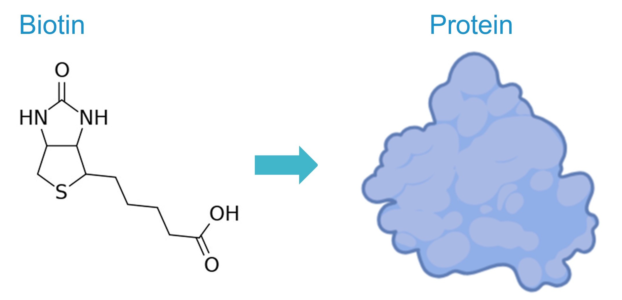
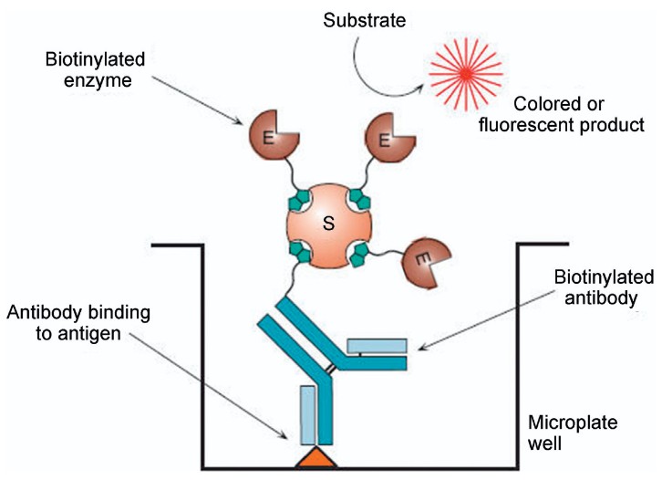 Figure 1. Biotin–streptavidin system. (Feng and Zhu, 2019)