We use cookies to understand how you use our site and to improve the overall user experience. This includes personalizing content and advertising. Read our
Privacy Policy
The complement system is a tightly regulated network of plasma proteins that bridges innate and adaptive immunity, mediating pathogen opsonization, inflammatory signaling, and direct cell lysis through the Membrane Attack Complex (MAC). Dysregulation of complement activation—whether through excessive activation, deficient regulation, or autoantibody-driven consumption—underlies a broad spectrum of autoimmune, inflammatory, infectious, and thrombotic disorders. Profacgen offers a comprehensive suite of Complement Component Assays designed to deliver quantitative, specific, and reproducible measurements of individual complement proteins and activation products. Our assays support biomarker discovery, diagnostic development, therapeutic monitoring, and mechanistic studies across classical, lectin, and alternative pathway biology.
What We Offer
Profacgen provides customizable and ready-to-use assay solutions for the precise quantification of complement components, split products, and regulatory factors. Our platform accommodates diverse sample types—including serum, plasma, cell culture supernatants, and tissue lysates—while maintaining strict specificity without cross-reactivity against related complement proteins.
Single-Analyte Quantification
Targeted assays for individual complement proteins (C3, C4, C5, C1q, Factor B, Factor H, Factor I, and others) using validated antibody pairs and calibrated reference standards.
Multiplex Complement Panels
Simultaneous measurement of multiple analytes in a single sample, enabling efficient profiling of pathway activation status and consumption patterns.
Activation Product Detection
Specialized assays for cleavage fragments and complexes (C3a, C3b, C5a, C4d, sTCC) to distinguish between native protein levels and pathway activity.
Custom Assay Development
Tailored assay design for novel targets, non-human species, or unique matrix requirements, with full validation support for GLP-compliant studies.
Assay Principle and Workflow
Our complement component assays are built on solid-phase immunoassay principles, leveraging high-affinity capture and detection antibodies for sensitive and specific quantification. The general workflow proceeds through the following stages:
Sample Preparation: Serum or plasma samples are collected under standardized conditions to minimize ex vivo complement activation. EDTA, citrate, or specialized protease inhibitor cocktails are used as appropriate for the target analyte.
Capture Phase: Microplate wells are coated with target-specific capture antibodies, enabling immobilization of the complement protein or activation product from the sample matrix.
Detection and Quantification: Bound analytes are detected using enzyme-conjugated or fluorophore-labeled secondary antibodies. Signal intensity is measured spectrophotometrically or fluorometrically and interpolated against a calibrated standard curve.
Data Analysis and Reporting: Concentrations are calculated using four- or five-parameter logistic regression. Results are reported with full QC metrics, including intra- and inter-assay precision, recovery, and linearity.
Service Capabilities
Profacgen's complement assay platform integrates multiple detection modalities and flexible formatting to match the analytical requirements of each project.
Capability
Description
Best Suited For
ELISA-Based Quantification
Sandwich and competitive ELISA formats with colorimetric, chemiluminescent, or fluorescent readouts. Detection limits typically in the pg/mL to ng/mL range.
Light-scattering measurement of immune complex formation for rapid, automated quantification of native complement protein concentrations.
Clinical diagnostics, routine monitoring of C3 and C4 levels
Multiplex Assays
Bead-based or planar array platforms enabling simultaneous detection of up to 20+ complement-related analytes per well.
Pathway profiling, drug mechanism studies, biomarker panel screening
Functional Hemolytic Assays
Cell-based assays measuring complement-mediated lysis of antibody-sensitized sheep erythrocytes (CH50) or rabbit erythrocytes (AH50) to assess total classical or alternative pathway activity.
Global pathway activity assessment, therapeutic efficacy evaluation
Supported Targets
Our validated assay menu covers key components across all activation pathways and regulatory nodes:
Central Components: C3, C4, C5
Initiation and Recognition: C1q, Mannose-Binding Lectin (MBL)
Figure 1. Three complement activation cascades—classical, lectin, and alternative pathways converge at C3 convertase formation. (Zhou et al., 2008)
Applications
Complement component assays serve critical roles across translational research, diagnostic development, and therapeutic monitoring:
Autoimmune Disease Monitoring: Quantification of C4d as a stable biomarker for antibody-mediated rejection in transplantation, and C3/C4 consumption patterns in systemic lupus erythematosus (SLE) flare assessment.
Thrombotic Microangiopathy Diagnosis: Measurement of CFH, Factor I, and C3 levels to differentiate atypical hemolytic uremic syndrome (aHUS), membranoproliferative glomerulonephritis (MPGN), and C3 glomerulopathy.
Age-Related Macular Degeneration (AMD): Genetic and biochemical analysis of CFH polymorphisms and circulating CFH levels to stratify risk and evaluate complement-targeting therapeutics.
Complement Therapeutic Development: Pharmacodynamic monitoring of anti-C5, anti-C3, and C1q inhibitors; assessment of target engagement and pathway modulation in preclinical and clinical samples.
Infectious Disease Research: Evaluation of complement consumption during sepsis, assessment of pathogen evasion strategies, and correlation of complement levels with disease severity.
Neurodegenerative Biomarker Studies: Profiling complement activation in Alzheimer's disease, multiple sclerosis, and other CNS disorders where synaptic pruning and neuroinflammation are complement-dependent.
Service Advantages
Pathway-Specific Specificity: Antibodies validated for discrimination between native proteins, cleavage products, and regulatory isoforms without cross-reactivity
Flexible Detection Platforms: ELISA, nephelometry, and multiplex formats tailored to throughput, sensitivity, and sample volume constraints
Pre-Validated Panels: Ready-to-deploy complement panels with established dynamic ranges and performance characteristics
GLP-Compatible Workflow: Assay validation, documentation, and QC protocols suitable for regulated preclinical and clinical studies
Multi-Species Capability: Assay formats available for human, mouse, rat, cynomolgus monkey, and other non-human primate samples
Integrated Data Interpretation: Expert support for pathway mapping, consumption pattern analysis, and biomarker correlation studies
Deliverables
Each complement assay project is delivered with comprehensive documentation and analytical support:
Quantitative analyte concentrations with unit-standardized reporting (µg/mL, ng/mL, or pg/mL as appropriate)
Calibrated standard curves with regression parameters and goodness-of-fit metrics
Full QC data including intra-assay CV, inter-assay CV, spike-recovery, and dilution linearity
Raw optical density or fluorescence values upon request
Method validation reports for GLP-compliant studies
Expert consultation on data interpretation and pathway-level insights
Case 1: C4d Biomarker Validation for Antibody-Mediated Rejection Monitoring in Renal Transplantation
Background:
A clinical diagnostics developer required a robust C4d quantification assay to support monitoring of antibody-mediated rejection (ABMR) in renal transplant recipients. Existing methods lacked the sensitivity to detect subtle C4d elevation in peritubular capillaries and serum, leading to delayed intervention and graft loss. The client needed a standardized assay with demonstrated correlation to Banff classification criteria and clinical outcomes.
Our Solution:
Profacgen developed a high-sensitivity ELISA for C4d detection in serum and paraffin-embedded tissue lysates, utilizing a neoantigen-specific capture antibody that distinguishes C4d from native C4. The assay was cross-validated against immunohistochemistry scoring and donor-specific antibody (DSA) profiles in a retrospective cohort of 120 transplant recipients. We established metrics for assay precision, linearity and stability that are aligned with the guidance of regulatory agencies on the qualification of biomarkers.
Final Results:
The validated C4d assay achieved a detection limit of 0.5 ng/mL with inter-assay CV below 8%, enabling reliable discrimination between stable graft function and subclinical ABMR. The client successfully incorporated the assay into a multi-analyte transplant monitoring panel, supporting prospective clinical validation and regulatory compliance.
Case 2: CFH Quantification Panel for Atypical Hemolytic Uremic Syndrome (aHUS) Patient Stratification
Background:
A biotechnology company developing a complement Factor H (CFH) fusion protein for aHUS treatment required a companion diagnostic strategy to identify patients with CFH deficiency or dysfunction. Existing nephelometric methods could not differentiate between functional and non-functional CFH variants, complicating patient selection and therapeutic monitoring.
Our Solution:
Profacgen established a dual-assay approach combining total CFH quantification by ELISA with functional CFH activity assessment through C3b deposition inhibition. We developed species-cross-reactive reagents to support parallel preclinical studies in cynomolgus monkey and human matrices. Custom reference standards were calibrated against purified wild-type and mutant CFH proteins to ensure accurate genotype-phenotype correlation.
Final Results:
The integrated CFH panel enabled precise stratification of aHUS patients into CFH-deficient, CFH-functional-variant, and normal cohorts. The companion diagnostic supported the client's IND submission and facilitated Phase I trial enrollment, reducing screen failure rates by 35% through prospective patient identification.
Q: What is the difference between measuring total C3 and C3a/C3b activation products?
A: Total C3 quantification reflects the combined concentration of native C3 and its cleavage products, providing information on overall protein abundance. In contrast, C3a and C3b measurements specifically indicate complement activation status, as these split products are only generated when the classical, lectin, or alternative pathway is actively engaged. For pathway activity assessment, activation product detection is essential.
Q: Which sample type is preferred for complement assays—serum or plasma?
A: The optimal sample type depends on the target analyte. EDTA plasma is generally preferred for activation products (C3a, C5a, sTCC) because EDTA chelates calcium and magnesium, halting ex vivo complement activation during collection. For native protein quantification (total C3, C4, C5), serum or citrate plasma may be acceptable. We provide detailed collection guidelines tailored to each assay format.
Q: Can complement assays distinguish between the classical, lectin, and alternative pathways?
A: Yes, through strategic analyte selection. C1q and C4d are primarily associated with classical and lectin pathway activation, while Factor B and Properdin are specific to the alternative pathway. C3 and C5 cleavage products are common to all three pathways. By profiling pathway-specific components, we can infer which activation route is predominantly engaged in a given sample.
Q: Are your complement assays suitable for monitoring complement inhibitor therapeutics?
A: Absolutely. Our assays are routinely used for pharmacodynamic monitoring of complement-targeting drugs, including anti-C5 antibodies (eculizumab, ravulizumab), C1q inhibitors, and C3-targeting agents. We can measure target engagement, free versus total drug levels, and residual pathway activity to support dose optimization and efficacy evaluation.
Q: What is the typical turnaround time for a complement panel project?
A: Standard turnaround for pre-validated single-analyte ELISAs is 2–3 weeks from sample receipt. Custom multiplex panel development typically requires 4–6 weeks for assay optimization and validation. Large-scale clinical or GLP-compliant studies are scheduled based on project scope and sample volume, with dedicated project management throughout.
Q: Do you offer complement assays for non-human species?
A: Yes. In addition to our extensively validated human complement assays, we have developed or can custom-develop assays for mouse, rat, cynomolgus monkey, and other species commonly used in preclinical research. Cross-reactivity testing and species-specific reference standards are incorporated into all non-human assay validations.
References:
Zhou X, Hu W, Qin X. The role of complement in the mechanism of action of rituximab for b-cell lymphoma: implications for therapy. The Oncologist. 2008;13(9):954-966. doi:10.1634/theoncologist.2008-0089
Online Inquiry
Fill out this form and one of our experts will respond to you within one business day.

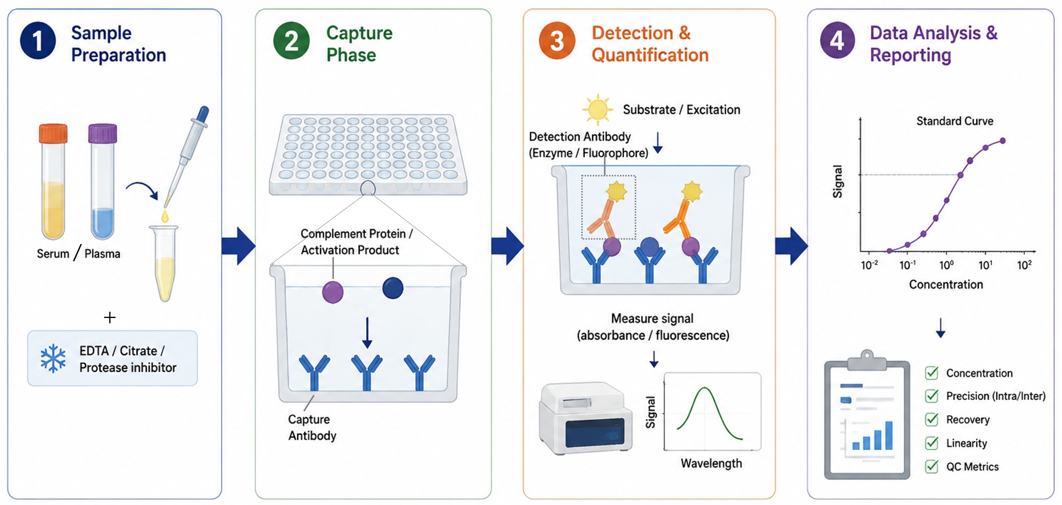
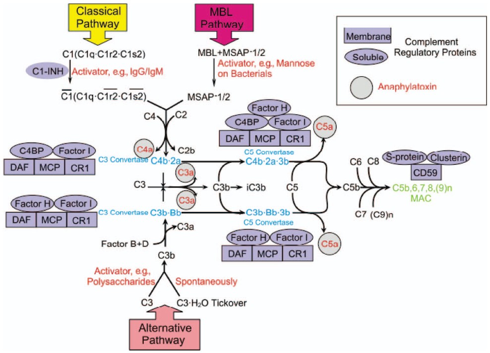 Figure 1. Three complement activation cascades—classical, lectin, and alternative pathways converge at C3 convertase formation. (Zhou et al., 2008)