We use cookies to understand how you use our site and to improve the overall user experience. This includes personalizing content and advertising. Read our
Privacy Policy
Bimolecular Fluorescence Complementation (BiFC) is a powerful imaging technology that enables the direct visualization of protein-protein interactions (PPIs) in their native cellular context with spatial and temporal precision. The method exploits a unique property of fluorescent proteins: when split into two non-fluorescent fragments and fused to potentially interacting proteins, the fragments reassemble into a functional fluorophore only when the target proteins come into close proximity—providing an unambiguous, real-time readout of where, when, and how strongly two proteins interact inside living cells.
Figure 1. BiFC assay to determine interactions of the CTPS 1-4. (Krämer et al., 2022)
BiFC has been successfully applied across a remarkable range of cell types and organisms, from E. coli and yeast to mammalian cells and whole plants. Unlike methods that require cell lysis or exogenous dyes, BiFC operates entirely within the living cell under conditions that closely approximate the physiological state, capturing transient interactions, weak associations, and the dynamic regulation of protein complexes by cellular signaling molecules. Profacgen offers a comprehensive BiFC service platform built on optimized split-fluorescent protein systems, experienced molecular biology expertise, and quantitative imaging capabilities to advance your protein interaction research.
Background: Visualizing the Invisible Interactome
Protein-protein interactions form the molecular circuitry of the cell. An estimated 130,000–650,000 distinct PPIs operate in human cells, governing virtually every biological process from DNA replication and transcription to signal transduction and immune defense. Mapping this "interactome" has been one of the grand challenges of molecular biology, driving the development of increasingly sophisticated detection technologies.
Traditional methods for studying PPIs—yeast two-hybrid, co-immunoprecipitation, and affinity chromatography—have generated invaluable data but share a common limitation: they measure interactions outside the native cellular environment. Cell lysis disrupts subcellular compartments, dilutes interaction partners, and removes the regulatory cues that modulate binding in vivo. Förster Resonance Energy Transfer (FRET) overcame this limitation by enabling live-cell detection, but its requirement for precise donor-acceptor orientation and close proximity (<10 nm) limits its applicability to well-characterized protein pairs.
BiFC represents a conceptual breakthrough by turning protein interaction itself into a fluorescent signal. The method builds on the discovery by Kerppola and colleagues that the β-barrel structure of GFP-family proteins contains permissive loop regions where foreign proteins can be inserted without abolishing fluorescence. By splitting the fluorescent protein at these loops and fusing each fragment to a candidate interacting protein, the cell becomes a self-reporting system: interaction produces light, observable by standard fluorescence microscopy. This simplicity, combined with the ability to detect interactions in any subcellular compartment without exogenous reagents, has made BiFC one of the most widely adopted tools in cell biology and drug discovery.
Figure 2. Schematic representation of the principle of the BiFC assay. (Kerppola, 2008)
Our BiFC Systems
Split-YFP (Enhanced Yellow Fluorescent Protein)
The gold-standard BiFC system for strong, stable protein interactions.
Split at residue 155 (YN: aa 1–154; YC: aa 155–238)
Bright fluorescence with low background interference
Fast maturation at 37 °C (suitable for mammalian systems)
Optimal for visualizing interaction subcellular localization
Lower fluorescence intensity minimizes detection of weak/transient interactions
Split-Venus
An improved YFP variant with enhanced folding and reduced maturation time.
Faster fluorophore maturation enables detection of transient interactions
Higher fluorescence intensity than standard YFP
Improved signal-to-background ratio at 37 °C
Ideal for mammalian cell and tissue imaging
Split-mCherry / Split-tdTomato
Red-fluorescent BiFC for spectral multiplexing with GFP-based assays.
Red emission enables co-localization with GFP-tagged organelle markers
Reduced phototoxicity compared to UV-excited fluorophores
Compatible with two-color BiFC (simultaneous detection of two PPIs)
Self-complementing GFP fragments for irreversible interaction detection.
Fragment 1–10 ( detector) complements fragment 11 (1–2 kDa tag)
The small fragment 11 can be genetically fused without disrupting protein function
Irreversible complementation traps even transient interactions
Excellent for screening libraries and mapping interaction networks
Service Workflow
Experimental Design Options
Design Feature
Options
Considerations
Fragment fusion position
N-terminal or C-terminal fusion of BiFC fragments to target proteins
N-terminal fusion may interfere with signal peptides or targeting sequences; C-terminal fusion may affect protein stability. Both should be tested when possible.
Expression system
Mammalian (HEK293, HeLa, CHO), plant (Nicotiana benthamiana), yeast (S. cerevisiae), bacteria (E. coli)
Choice depends on the native expression context of the proteins, post-translational modification requirements, and imaging infrastructure.
Consider spectral compatibility with other fluorescent markers, brightness at the experimental temperature, and the kinetic stability of the interaction.
Microscopy provides subcellular localization; flow cytometry enables quantitative comparison across large populations; plate readers offer cost-effective screening.
Controls
Non-interacting protein pairs, single-fragment transfections, interaction-disrupting mutations
Essential for distinguishing true interactions from spontaneous fragment complementation and for estimating interaction strength.
Applications
In-cellulo protein-protein interaction mapping: Visualize where two proteins interact within the cell—nucleus, cytoplasm, membrane, or specific organelles—providing spatial context unavailable from biochemical methods
Interaction dynamics and regulation: Monitor how protein interactions change in response to hormones, small molecules, signaling events, or environmental stimuli in real time
Interaction strength quantification: Compare fluorescence intensity across different protein pairs and mutants to rank interaction affinities and identify critical interface residues
Drug target validation: Confirm that a candidate therapeutic modulates the intended protein interaction by observing changes in BiFC signal upon compound treatment
Protein complex topology: Map the arrangement of subunits within multi-protein assemblies using pairwise BiFC combinations
Competition and displacement assays: Screen for compounds that disrupt a specific protein interaction by measuring BiFC signal reduction
Why Choose Profacgen?
Multiple Fluorescent Systems: YFP, Venus, mCherry, tdTomato, and large-segment GFP for flexible experimental design.
Living Cell Context: Observe interactions under physiological conditions, preserving native folding, post-translational modifications, and subcellular localization.
No Structural Information Required: BiFC works without prior knowledge of protein structures, making it ideal for exploratory interaction screens.
Subcellular Resolution: Fluorescence microscopy pinpoints the exact location of interactions within the cell.
Quantitative Options: Flow cytometry and plate reader formats enable high-throughput quantitative screening and interaction strength ranking.
Comprehensive Validation: Co-immunoprecipitation and immunofluorescence validation available as orthogonal confirmation.
Representative Case Studies
Case 1: Mapping the Subcellular Interaction Network of a Viral Replication Complex
Background:
A virology research group studying SARS-CoV-2 replication needed to determine which viral non-structural proteins (nsps) interacted with each other and where these interactions occurred within infected cells. Traditional co-IP had identified 12 binary interactions but provided no spatial or temporal information.
Our Solution:
Profacgen designed a systematic BiFC screen using the split-Venus system in HEK293 cells. All 66 pairwise combinations of 12 nsps were tested with both N-terminal and C-terminal fragment fusions. Confocal microscopy was used to visualize fluorescence patterns, and flow cytometry provided quantitative fluorescence intensity values for interaction strength ranking.
Final Results:
BiFC confirmed all 12 previously reported interactions and discovered 8 novel ones. Strikingly, nsp3-nsp4 interaction was exclusively localized to perinuclear puncta consistent with ER-derived replication organelles, while nsp7-nsp8 interaction was distributed diffusely in the cytoplasm before concentrating at replication sites 8 hours post-transfection. Mutational analysis of nsp3 identified a coiled-coil domain (residues 364–390) essential for nsp4 recruitment. These findings were published in a high-impact virology journal and informed subsequent antiviral drug design targeting the nsp3-nsp4 interface.
Case 2: Quantitative BiFC Screen Identifies Disruptors of the MDM2-p53 Oncoprotein Interaction
Background:
An oncology drug discovery program sought to identify small molecules that disrupt the MDM2-p53 protein-protein interaction—a validated cancer target where MDM2 ubiquitinates and degrades the tumor suppressor p53. Existing assays required cell lysis or expensive reagents, limiting throughput.
Our Solution:
Profacgen developed a quantitative BiFC assay using split-YFP: p53 was fused to YN and MDM2 to YC. The assay was optimized in HCT116 cells and validated with the known inhibitor nutlin-3a (positive control) and a non-binding MDM2 mutant (negative control). Flow cytometry provided a quantitative readout amenable to 96-well screening format. A library of 5,000 FDA-approved drugs and natural products was screened.
Final Results:
The BiFC assay achieved a Z′ of 0.71 with nutlin-3a producing a 65% signal reduction. Screening identified 14 compounds that reduced the BiFC signal by >40%. Four compounds were confirmed by orthogonal co-IP assays and showed dose-dependent p53 stabilization and transcriptional activation of p21 and BAX. The lead compound, a repurposed cardiac glycoside, exhibited an IC50 of 2.3 μM and is now in preclinical development for TP53-wildtype colorectal cancer.
Q: What is the difference between BiFC and FRET for studying protein interactions?
A: BiFC and FRET are complementary live-cell interaction detection methods with distinct advantages. BiFC produces a strong, irreversible fluorescent signal upon interaction, making it highly sensitive for detecting weak or transient interactions and ideal for determining subcellular localization. However, the irreversible complementation means BiFC cannot track the dissociation of an interaction in real time. FRET, by contrast, is fully reversible and can monitor association and dissociation kinetics, but it requires precise donor-acceptor orientation and close proximity (<10 nm), producing weaker signals that are more challenging to detect. For many projects, BiFC and FRET are used together: BiFC to confirm and localize interactions, and FRET to study their dynamics.
Q: Can BiFC detect transient protein interactions?
A: BiFC can detect transient interactions, but with important caveats. Because fluorescent protein fragment complementation is effectively irreversible on physiological timescales, a transient interaction that brings the fragments together for even a few seconds can produce a stable fluorescent signal that persists after dissociation. This "molecular memory" makes BiFC highly sensitive for detecting transient interactions but prevents measurement of their true kinetic parameters. For quantifying transient interaction kinetics, we recommend complementary methods such as FRET or BRET. The split-Venus system, with its faster maturation, provides the best balance for detecting transient interactions while minimizing artifact accumulation.
Q: Does the fluorescent protein fragment fusion interfere with protein function?
A: In most cases, BiFC fragments (typically 80–120 amino acids) can be fused to target proteins without disrupting function, particularly when fused at flexible termini. However, every protein pair is unique. We recommend testing both N-terminal and C-terminal fusions and validating that the fusion proteins retain their expected subcellular localization, expression level, and known interaction partners. As part of our standard workflow, we perform control experiments including immunoblotting for expression verification and co-immunoprecipitation to confirm that the BiFC signal reflects a genuine interaction.
Q: What is the typical timeline for a BiFC project?
A: A standard BiFC project takes 4–6 weeks: plasmid construction and sequence verification (1–2 weeks), cell transfection and expression optimization (1 week), confocal imaging and qualitative analysis (1–2 weeks), and data interpretation and report preparation (1 week). Quantitative flow cytometry projects add 1–2 weeks for assay optimization and statistical validation. Large-scale pairwise screens (e.g., 50+ combinations) require 8–12 weeks.
Q: Can BiFC be used for high-throughput screening?
A: Yes. By coupling BiFC with flow cytometry or automated fluorescence plate readers, BiFC can be adapted for medium- to high-throughput screening. The split-GFP large-segment system is particularly well-suited for screening because its irreversible complementation produces a robust signal that is easily detected by standard instruments. We have successfully used BiFC to screen compound libraries of up to 10,000 molecules for interaction disruptors. For very large libraries (>50,000 compounds), we recommend a tiered approach with an initial biochemical screen followed by BiFC validation in cells.
Q: How do you control for false positives from spontaneous fragment complementation?
A: Every BiFC experiment includes multiple negative controls: (1) each fragment expressed alone to assess spontaneous fluorescence; (2) a non-interacting protein pair (e.g., fragments fused to proteins known not to interact) to establish background levels; (3) interaction-disrupting point mutations in one binding partner to confirm signal specificity; and (4) where possible, a competing unlabeled interaction partner to demonstrate saturation. A positive BiFC result is reported only when the test signal significantly exceeds all negative controls (typically >3-fold above background with p < 0.01).
References:
Krämer M, Dörfer E, Hickl D, Bellin L, Scherer V, Möhlmann T. Cytidine triphosphate synthase four from arabidopsis thaliana attenuates drought stress effects. Front Plant Sci. 2022;13:842156. doi:10.3389/fpls.2022.842156
Kerppola TK. Bimolecular fluorescence complementation (BiFC) analysis as a probe of protein interactions in living cells. Annu Rev Biophys. 2008;37(1):465-487. doi:10.1146/annurev.biophys.37.032807.125842
Online Inquiry
Fill out this form and one of our experts will respond to you within one business day.

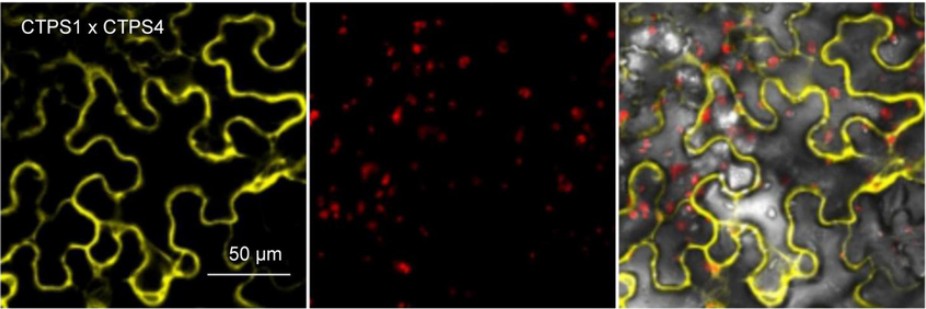 Figure 1. BiFC assay to determine interactions of the CTPS 1-4. (Krämer et al., 2022)
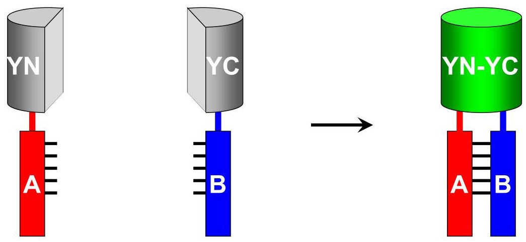 Figure 2. Schematic representation of the principle of the BiFC assay. (Kerppola, 2008)