Single-Cell Enzyme Assays
Cellular heterogeneity underlies the functional diversity of tissues, the progression of disease, and the variable response of patients to therapeutic intervention. While bulk biochemical assays average signals across millions of cells, obscuring rare subpopulations and masking critical biological differences, single-cell enzyme assays resolve enzyme activity at the level of the individual cell. This resolution is essential for understanding how genetic, epigenetic, and microenvironmental factors generate phenotypic diversity within apparently homogeneous populations. Profacgen provides advanced single-cell enzyme assay services that combine microfluidic confinement, fluorescence imaging, and high-throughput flow cytometry to quantify enzyme activity, signaling dynamics, and metabolic states in single cells with precision and reproducibility.
Single-Cell Detection Technologies
Single-cell enzyme activity analysis is a real-time, in situ, minimally invasive detection technology that quantifies signaling molecules, biomarkers, and enzyme activities at subcellular resolution. At present, single-cell enzyme analysis is performed primarily through two complementary approaches: image-based analyses and flow-based systems. Profacgen integrates both modalities to match the biological question, throughput requirements, and sensitivity demands of each project.
Microfluidics
Microfluidic devices enable the precise manipulation of picoliter-to-nanoliter volumes, allowing single cells and reagents to be confined within discrete microdroplets or channel-bound chambers. This confinement eliminates cross-contamination, reduces reagent consumption by orders of magnitude, and accelerates reaction kinetics through enhanced molecular crowding.
The microfluidic workflow Profacgen employs proceeds as follows:
- Cell and Substrate Loading: A cell suspension and a fluorogenic peptide substrate are introduced into the microfluidic device through separate inlet channels. The substrate contains a peptide sequence specific to the target enzyme, flanked by a fluorescent donor group at one terminus and a quencher acceptor at the other.
- Droplet Generation: The two aqueous phases meet at a flow-focusing junction and are segmented into monodisperse microdroplets by a continuous oil phase. Statistical dilution ensures that the majority of droplets contain either zero or one cell (Poisson distribution), with droplets containing single cells selected for downstream analysis.
- Incubation and Reaction: Within each droplet, the cell is lysed or permeabilized, releasing intracellular enzymes that cleave the peptide substrate. Enzymatic cleavage separates the fluorophore from the quencher, restoring fluorescence emission proportional to enzyme activity.
- Fluorescence Detection: Droplets flow through a detection region under the drive of the continuous phase, where a fluorescence microscope or integrated photodetector records the signal from each droplet individually.
- Data Analysis: Fluorescence intensities are mapped to enzyme activity through calibration curves generated with recombinant enzyme standards. Single-cell activity distributions are plotted to reveal population heterogeneity, identify rare high-activity subpopulations, and correlate activity with co-encapsulated biomarkers.
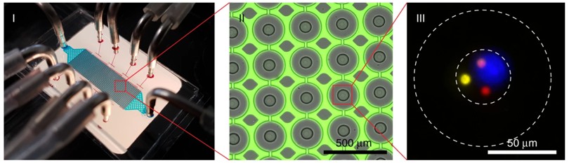 Figure 1. Microfluidic single-cell enzyme assay: droplet generation, encapsulation, and fluorescence detection.
Fluorescence Imaging
Image-based single-cell enzyme analysis is performed using high-resolution fluorescence microscopy in combination with fluorogenic or FRET (Förster Resonance Energy Transfer) substrates. This approach preserves spatial information, enabling the mapping of enzyme activity within subcellular compartments (e.g., cytosol, nucleus, mitochondria, lysosomes).
Key imaging modalities include:
- Wide-field fluorescence microscopy: Rapid acquisition of large cell populations for statistical heterogeneity analysis
- Confocal laser scanning microscopy: Optical sectioning for three-dimensional activity mapping with minimal out-of-focus blur
- Total internal reflection fluorescence (TIRF) microscopy: Evanescent wave excitation restricted to the basal ~100 nm of adherent cells, ideal for membrane-proximal enzyme studies
- FRET biosensors: Genetically encoded or synthetic FRET probes report enzyme activity through ratiometric changes in donor/acceptor fluorescence, enabling real-time kinetic monitoring in living cells
Early single-cell analyses relied on oil-layer confinement to isolate cell lysates within nanoliter droplets on microscope slides. Profacgen has transitioned to modern microfluidic devices that offer superior throughput, reproducibility, and compatibility with automated liquid handling.
Droplet-Based Systems
Droplet-based microfluidics represents a convergent platform that combines the compartmentalization of microfluidics with the scalability of flow-based detection. Monodisperse aqueous droplets suspended in a biocompatible fluorinated oil serve as independent picoliter reaction vessels, each harboring a single cell and its substrate cocktail. Droplets are generated at kilohertz frequencies, incubated in delay lines, and interrogated by fluorescence-activated droplet sorting (FADS) or imaging flow cytometry. This platform achieves throughputs of thousands of cells per second while retaining the resolution of single-cell analysis.
Instrumentation parameters optimized for droplet-based enzyme assays include:
- Droplet diameter: 20–100 µm (corresponding to 4–500 pL), matched to cell size and reagent cost considerations
- Generation frequency: Up to 10 kHz, enabling million-cell experiments within hours
- Fluorescence channels: Green (FITC, Alexa Fluor 488), red (Cy3, Alexa Fluor 555), and near-infrared (Cy5) for multiplexed enzyme and viability staining
- Sorting capability: Dielectrophoretic or acoustic sorting of droplets based on fluorescence threshold for downstream recovery and validation
Flow Cytometry
Flow-based systems remain the gold standard for high-throughput single-cell measurements. Flow cytometric analyses employ fluorescent antibodies, fluorogenic substrates, or phospho-specific probes to quantify enzyme (or phosphoenzyme) concentration as a proxy for enzyme activity. Modern instruments equipped with 20+ parameter panels enable simultaneous profiling of multiple enzymatic pathways, surface markers, and viability states in single cells. Profacgen supports both conventional fluorescence-activated cell sorting (FACS) and spectral flow cytometry for complex, multi-color experiments.
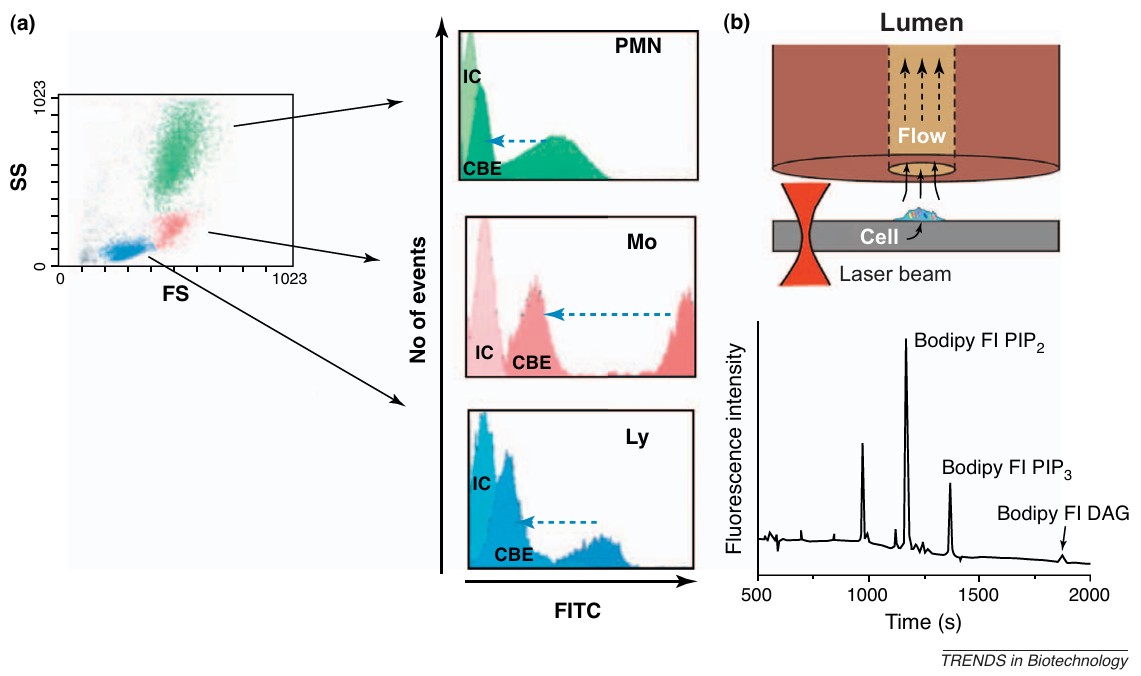 Figure 2. Flow- and separation-based methods for single-cell enzyme assays. (Kovarik and Allbritton, 2011)
Inquiry
Applications
Single-cell enzyme assays unlock biological insights that are inaccessible to population-averaged methods:
- Cellular heterogeneity: Resolution of enzyme activity distributions within clonal cell lines, primary tissues, and patient-derived samples reveals subpopulations with distinct metabolic or signaling states. This heterogeneity underlies drug resistance, stem cell maintenance, and developmental lineage decisions.
- Immune profiling: Single-cell phospho-flow cytometry maps kinase and phosphatase activities in immune cell subsets (T cells, B cells, NK cells, macrophages) under resting, activated, and exhausted states. These data inform immunotherapy design, biomarker discovery, and autoimmune disease monitoring.
- Cancer metabolism: Tumors are metabolically heterogeneous ecosystems in which rare cells with elevated glycolytic or oxidative enzyme activity drive relapse and metastasis. Single-cell assays identify these aggressive subpopulations and characterize their metabolic dependencies for targeted therapeutic intervention.
- Drug response prediction: Correlating single-cell enzyme activity with drug sensitivity profiles enables the identification of predictive biomarkers and the stratification of patient populations for personalized therapy.
- Stem cell and developmental biology: Tracking enzyme activity dynamics during differentiation reveals lineage-commitment checkpoints and the epigenetic mechanisms that stabilize cell fate.
Advantages Over Bulk Assays
- Resolution of Cellular Heterogeneity: Bulk assays average signals across millions of cells, masking rare but biologically critical subpopulations; single-cell assays capture the full distribution
- Minimal Sample Requirements: Microfluidic and droplet-based formats analyze enzyme activity in picoliter volumes, conserving precious clinical specimens and rare cell types
- Spatial and Temporal Information: Imaging-based approaches map subcellular enzyme localization and track activity dynamics in real time, impossible in lysate-based bulk methods
- Multiplexed Profiling: Simultaneous quantification of multiple enzymes, signaling nodes, and surface markers in the same cell reveals pathway crosstalk and phenotype–function relationships
- Direct Correlation with Genotype: Single-cell sequencing can be paired with enzyme activity measurements to link genetic mutations, copy number variations, or transcriptomic states to functional phenotypes
- High-Throughput Capability: Droplet microfluidics and flow cytometry analyze thousands to millions of cells per experiment, combining single-cell resolution with statistical power
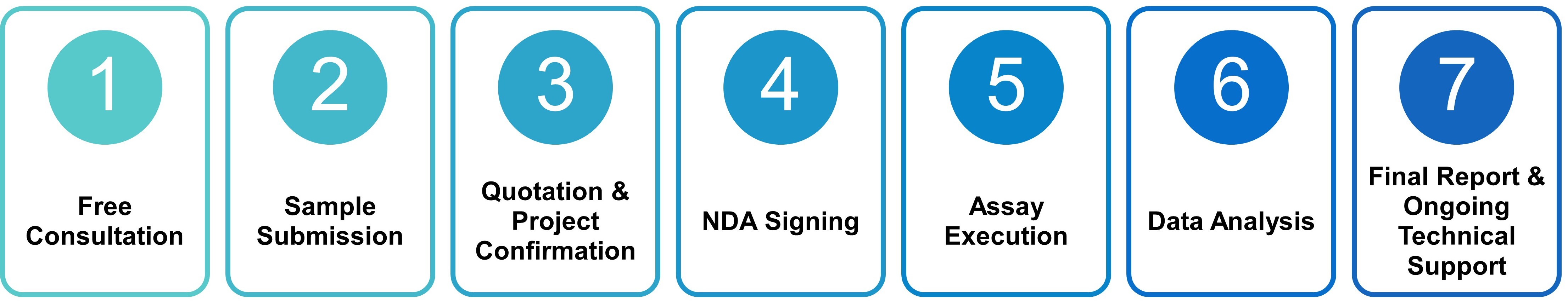
Workflow of Single-Cell Enzyme Assay
Why Choose Profacgen?
Profacgen combines cutting-edge instrumentation, rigorous experimental design, and client-focused project management to deliver single-cell enzyme assay data of the highest quality:
- Short turnaround, high quality: Efficient project timelines with comprehensive experimental schemes and scientifically rigorous, fair, and accurate test reports
- Advanced instrumentation: Access to international-grade microfluidic fabrication facilities, high-content imaging platforms, and spectral flow cytometers housed in dedicated research laboratories
- Confidentiality and privacy: All projects are protected by confidentiality agreements with strict data security protocols and customer privacy safeguards
- 24/7 online support: Dedicated project managers and scientific consultants available around the clock to address technical questions, review interim data, and adapt experimental plans
- Integrated workflow: Seamless connection from single-cell enzyme assays to downstream genomic profiling, proteomics, and functional validation studies
Representative Case Studies
Case 1: Single-Cell Phosphatase Profiling in Drug-Resistant Cancer Cells
Background:
An oncology research team observed heterogeneous drug responses within a supposedly homogeneous triple-negative breast cancer cell line. Bulk phosphatase assays failed to explain the persistence of a resistant subpopulation following targeted therapy.
Our Solution:
Profacgen employed droplet-based microfluidics to encapsulate single cells with a fluorogenic phosphatase substrate specific to the pro-survival phosphatase PP2A. Following lysis and reaction, droplets were analyzed by fluorescence-activated droplet sorting to map the single-cell phosphatase activity distribution.
Final Results:
The assay revealed a bimodal activity distribution with a rare high-PP2A subpopulation (~3% of cells) that correlated with elevated AKT signaling and doxorubicin resistance. Targeting this subpopulation with a PP2A inhibitor sensitized the population to chemotherapy, providing a rational combination strategy.
Case 2: Immune Cell Kinase Heterogeneity by Spectral Flow Cytometry
Background:
A biotechnology company developing a JAK inhibitor for autoimmune disease required single-cell resolution of JAK-STAT pathway activation across peripheral blood mononuclear cell (PBMC) subsets to understand on-target and off-target effects.
Our Solution:
Profacgen designed a 16-color spectral flow cytometry panel combining surface markers (CD3, CD4, CD8, CD14, CD19, CD56) with phospho-specific antibodies against pSTAT1, pSTAT3, pSTAT5, and pERK1/2. PBMCs from healthy donors were stimulated with IFN-γ or IL-6 in the presence or absence of the JAK inhibitor.
Final Results:
The assay resolved cell-type–specific JAK inhibition profiles: CD14+ monocytes showed the greatest pSTAT1 suppression (IC50 = 45 nM), while CD8+ T cells were less sensitive (IC50 = 320 nM). These data guided dose selection to maximize efficacy while preserving T-cell function.
Case 3: Microfluidic Single-Cell Glycolytic Enzyme Analysis in Tumor Biopsies
Background:
A pharmaceutical sponsor sought to identify metabolic subpopulations within patient-derived xenograft (PDX) tumor biopsies that might drive resistance to a glycolysis-targeting agent in clinical development.
Our Solution:
Profacgen developed a microfluidic assay encapsulating single dissociated tumor cells with a fluorogenic lactate dehydrogenase (LDH) substrate and a viability dye. Droplets were imaged by high-speed fluorescence microscopy, and LDH activity was quantified per cell.
Final Results:
Single-cell analysis revealed a metabolically distinct subpopulation with 5-fold elevated LDH activity that was enriched post-treatment and correlated with elevated hypoxia-inducible factor-1α (HIF-1α) expression. These cells were subsequently confirmed to drive tumor relapse in mouse models.
Get a Project Assessment
Frequently Asked Questions (FAQs)
Q: What is the difference between single-cell and bulk enzyme assays?
A: Bulk enzyme assays measure the average activity of millions of cells in a pooled lysate, obscuring cell-to-cell variability and rare subpopulations. Single-cell assays resolve enzyme activity in individual cells, revealing heterogeneity that is critical for understanding drug resistance, immune responses, and developmental processes.
Q: What sample types are compatible with single-cell enzyme assays?
A: We routinely analyze adherent and suspension cell lines, primary cells (PBMCs, tumor-infiltrating lymphocytes, fibroblasts), stem cell cultures, and dissociated tissue biopsies. Sample viability and single-cell suspension quality are the primary determinants of assay success; our team provides guidance on optimal dissociation and handling protocols.
Q: How many cells are needed for a single-cell enzyme assay?
A: Microfluidic and droplet-based assays require 104–105 cells for robust statistical analysis. Flow cytometry-based assays can analyze 106–107 cells per run. We recommend consulting with our team during project planning to determine the optimal cell number based on the expected subpopulation frequency and desired statistical power.
Q: Can single-cell enzyme assays be combined with genomics or transcriptomics?
A: Yes. Profacgen supports multi-omics integration through sequential or parallel workflows. For example, cells can be sorted by FADS based on enzyme activity, followed by single-cell RNA sequencing (scRNA-seq) or targeted qPCR to correlate functional phenotypes with gene expression states. Alternatively, imaging-based assays can be paired with in situ hybridization for spatial transcriptomics.
Q: What is the typical turnaround time for a single-cell enzyme assay project?
A: Standard projects using validated substrates and cell types are completed within 3–4 weeks. Custom assay development (novel substrate design, microfluidic chip fabrication, or panel optimization) extends the timeline to 6–8 weeks. Expedited services are available for time-critical studies.
Q: How do you ensure data quality and reproducibility in single-cell assays?
A: We implement rigorous quality control at every stage: cell viability assessment, droplet monodispersity verification, fluorescence calibration with recombinant enzyme standards, positive and negative control inclusion, and replicate analysis across independent microfluidic runs or flow cytometry acquisitions. All data are analyzed with validated statistical pipelines and reported with confidence intervals and technical replicate statistics.
References:
- Kovarik ML, Allbritton NL. Measuring enzyme activity in single cells. Trends Biotechnol. 2011;29(5):222–230. doi:10.1016/j.tibtech.2011.01.003