
Profacgen offers high-precision Biomembrane Force Probe (BFP) services for single-molecule mechanical characterization of receptor-ligand interactions. This biophysical technique quantifies individual molecular bonds with piconewton sensitivity across a wide force range (0.1 pN to 1 nN) and loading rates (1–106 pN/s), making it ideally suited for studying cellular adhesion, signaling, and immune recognition under physiologically relevant conditions.
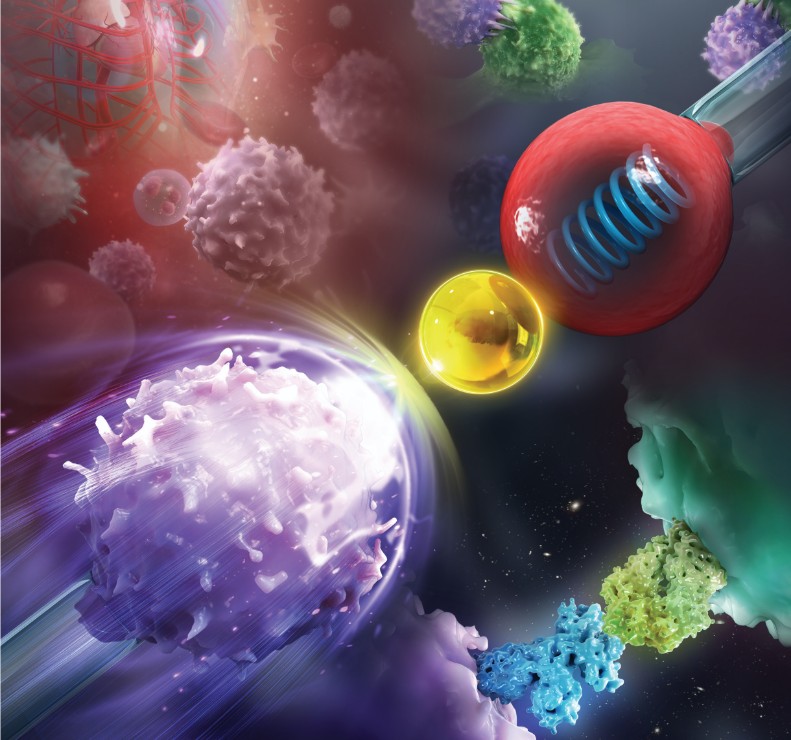
Our BFP platform employs a classic configuration: a red blood cell aspirated by a micropipette serves as a force sensor, with a ligand-coated glass microbead attached to the cell apex to probe interactions with target cells (e.g., platelets, leukocytes, or engineered lines). By monitoring erythrocyte deformation under calibrated tension, we precisely measure bond rupture forces, kinetic rates, and mechanical regulation of receptor function—all at the single-molecule level. Profacgen delivers robust, reproducible BFP data to support your drug discovery, receptor biology, and mechanobiology research.
Biological systems are fundamentally mechanical. Cells exert and sense forces through specialized receptor-ligand bonds that translate physical stimuli into biochemical signals—a process termed mechanotransduction. The immune system relies on mechanical forces to discriminate between self and non-self: T-cell receptors pull on peptide-MHC complexes to trigger signaling cascades; platelets sense shear forces through integrin αIIbβ3 to initiate clot formation; and leukocytes roll along vessel walls via selectin-mediated catch bonds that strengthen under force.
Understanding these force-dependent interactions requires tools that can apply and measure forces at the molecular scale. Atomic force microscopy (AFM) and optical tweezers were early pioneers, but each had limitations: AFM lacked the dynamic range for cellular studies, and optical tweezers required specialized optical setups. The Biomembrane Force Probe, developed in the early 2000s by the Zhu laboratory, addressed these limitations by using the red blood cell as a natural spring—exquisitely sensitive, biocompatible, and mechanically well-characterized.
The dual Biomembrane Force Probe (dual-BFP), developed more recently, represents a significant advance by incorporating two independent force probes. Probe I applies a precisely controlled mechanical stimulus while Probe II simultaneously monitors the molecular response—enabling the dissection of signal crosstalk between distinct receptor systems on the same cell. This capability has opened new frontiers in understanding how cells integrate multiple mechanical and chemical cues to make functional decisions.
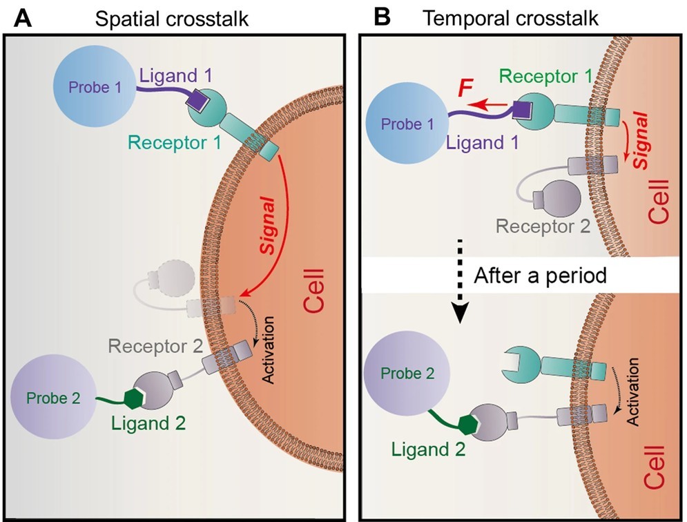 Figure 1. Concept of the dual biomembrane force probe (dBFP). (Ju et al., 2017)
| Parameter | Specification |
|---|---|
| Force range | 0.1 pN – 1 nN |
| Loading rate range | 1 – 106 pN/s |
| Mechanical resolution | ~1 pN |
| Off-rate detection limit | 10−3 s−1 |
| Affinity dynamic range | 10 mM – 1 nM (dissociation constant KD) |
| Temporal resolution | Millisecond (sub-millisecond with fast force clamp) |
| Probe configurations | Single-BFP and dual-BFP (simultaneous dual-receptor interrogation) |
| Target cell types | Platelets, leukocytes, endothelial cells, engineered cell lines, primary patient cells |
Traditional Single-BFP
Single-probe force spectroscopy for receptor-ligand bond characterization.
Dual-BFP
Two independent probes for mechanical stimulation and molecular response monitoring.
Multi-Ligand Presentation
Investigate cooperative binding of multiple receptor-ligand species.
Live-Cell Stimulation
Single-cell stimulation without paracrine signaling interference.
Background:
A biopharmaceutical company developing antithrombotic antibodies targeting integrin αIIbβ3 needed to understand how mechanical force—present in flowing blood—affected the binding properties of their lead candidates. Standard SPR measurements had identified three candidates with similar equilibrium affinities, but in vivo efficacy differed dramatically.
Our Solution:
Profacgen employed the dual-BFP platform to characterize all three antibody candidates under mechanical loading conditions mimicking physiological shear (1–100 pN/s). For each candidate, we measured the force-dependent off-rate, the rupture force distribution, and the bond lifetime as a function of applied force to classify each interaction as a slip bond, catch bond, or ideal bond.
Final Results:
Candidate A formed a catch bond with αIIbβ3—its lifetime increased 3-fold when force increased from 5 to 20 pN, then decreased at higher forces. This mechanical fingerprint predicted effective antithrombotic activity under arterial shear. Candidates B and C formed conventional slip bonds with monotonically decreasing lifetimes under force. In vivo arterial thrombosis models confirmed that only Candidate A significantly reduced thrombus formation (78% reduction vs. 12% and 8% for B and C). The force spectroscopy data were incorporated into the regulatory submission as a mechanistic rationale for clinical dosing.
Background:
A cell therapy company developing tumor-infiltrating lymphocyte (TIL) therapies observed that T-cell clones specific for certain neoantigens mediated potent tumor killing in vitro but failed to persist in vivo. They hypothesized that the mechanical binding properties of the T-cell receptor (TCR)–peptide-MHC (pMHC) interaction might determine T-cell activation threshold and persistence.
Our Solution:
Profacgen used the BFP to measure the force-lifetime relationship for 8 neoantigen-specific TCRs interacting with their cognate pMHC complexes. For each TCR, we constructed force vs. lifetime curves, identified the peak force for maximum bond lifetime, and correlated these mechanical parameters with T-cell activation markers (CD69, IL-2, IFN-γ) measured by flow cytometry.
Final Results:
TCRs with an optimal catch bond profile (peak lifetime at 10–15 pN) triggered robust T-cell activation and IL-2 production, while TCRs with monotonic slip bonds or catch bonds peaking at >25 pN showed diminished signaling. This mechanical optimization principle guided the selection of the top 3 TCR clones for clinical manufacturing. In the Phase I trial, patients receiving TIL products enriched for optimal catch-bond TCRs showed a 60% objective response rate vs. 25% in historical controls.
References:
Fill out this form and one of our experts will respond to you within one business day.