
Fluorescence polarization (FP) is a powerful biophysical technique for studying the binding and interactions of molecules in solution. Unlike fluorescence intensity measurements, which quantify the total amount of emitted light, FP specifically measures the anisotropy of emitted light—the degree to which fluorescent molecules maintain their orientation during the excited-state lifetime. When a small fluorescent tracer binds to a larger molecule, its rotational diffusion slows dramatically, resulting in an increase in polarization that can be detected with high precision and sensitivity.
This fundamental principle makes FP an exceptionally versatile platform for quantifying protein-protein interactions, protein-DNA interactions, enzyme kinetics, and small-molecule binding. The assay is homogeneous (no wash steps required), non-radioactive, ratiometric (minimizing interference from fluorescence quenchers and colorimetric compounds), and fully miniaturizable to 384- and 1536-well plate formats—making it one of the most widely adopted technologies in high-throughput screening (HTS) campaigns for drug discovery. Profacgen provides a comprehensive fluorescence polarization platform with multiple fluorescent dyes, an upgraded detection system that dramatically reduces false-positive rates, and expert assay design to accelerate your research.
The theoretical foundation of fluorescence polarization was laid in 1926 by the French physicist Jean-Baptiste Perrin, who described how the rotational Brownian motion of fluorophores depolarizes emitted light. Small molecules tumble rapidly in solution, randomizing their orientation before fluorescence emission and yielding low polarization values. Large molecules rotate slowly, maintaining their alignment with the excitation polarization and producing high polarization signals. The magnitude of this difference is quantitatively related to molecular size, temperature, viscosity, and excited-state lifetime.
FP remained primarily a biophysical curiosity until the 1990s, when advances in plate reader instrumentation and fluorescent probe chemistry transformed it into a practical drug discovery tool. The landmark application came in 1995 with the development of FP-based kinase assays by scientists at PanVera, who demonstrated that FP could reliably detect the binding of fluorescently labeled ATP analogs to kinase active sites. Since then, FP has been adopted across virtually every target class in drug discovery, including GPCRs, nuclear receptors, proteases, kinases, and phosphatases.
Today, FP is one of the "big three" homogeneous assay technologies alongside time-resolved FRET (TR-FRET) and AlphaScreen, each with distinct strengths. FP's advantages include a simple readout requiring only standard fluorescence polarization-capable plate readers, no need for specialized donor-acceptor pairs, insensitivity to assay volume and meniscus effects, and compatibility with colored compounds that would quench or absorb light in intensity-based assays. Profacgen's FP platform builds on this legacy, incorporating modern dye chemistry, optimized detection protocols, and rigorous quality control to deliver reliable, publication-ready data.
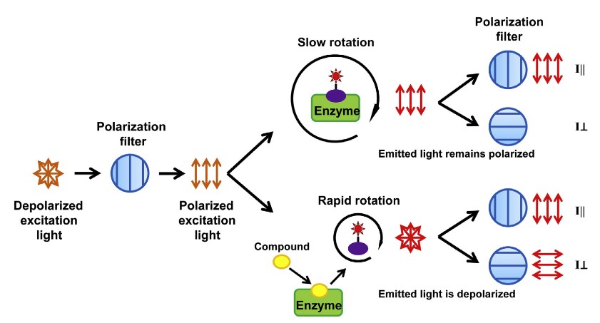 Figure 1. Principle of fluorescence polarization assay. (Zeng and Xu, 2015)
Non-Radioactive
Eliminates the safety, regulatory, and waste disposal burdens associated with 3H, 125I, and 32P labeling, while maintaining comparable sensitivity for most applications.
Homogeneous (“Mix and Read”)
No wash, separation, or transfer steps required. Reagents and sample are simply combined and read, minimizing handling time, pipetting errors, and assay variability.
Ratiometric Readout
The polarization value is a ratio of parallel and perpendicular fluorescence intensities, making it inherently insensitive to fluorescence quenchers, colored compounds, and minor pipetting volume variations.
Miniaturizable
Robust signal-to-background ratios enable assay miniaturization to 384-well (20–50 μL) and 1536-well (5–10 μL) formats, dramatically reducing reagent costs and expanding screening capacity.
| Service Category | Description | Typical Readout |
|---|---|---|
| Protein-Protein Interaction Assay | Quantify the binding affinity between two proteins by monitoring the change in polarization when a fluorescently labeled protein binds its unlabeled partner. Suitable for direct PPIs, domain-domain interactions, and competitive displacement. | KD (nM–μM); IC50 for competitive inhibitors |
| Protein-DNA/RNA Interaction Assay | Measure transcription factor–DNA binding, nucleic acid hybridization, or aptamer-target recognition using fluorescently labeled oligonucleotides as tracers. | KD, binding stoichiometry, sequence specificity |
| Enzyme Kinetic Assay | Monitor enzyme activity through product formation (increasing polarization as small substrate is converted to large product) or substrate depletion. Applicable to kinases, phosphatases, proteases, methyltransferases, and glycosyltransferases. | kcat, KM, kcat/KM; inhibitor IC50 |
| Small-Molecule Binding Assay | Identify and characterize small-molecule ligands through competitive displacement of a fluorescent tracer from the target binding site. The primary assay format for HTS hit identification. | % inhibition; IC50; binding site competition analysis |
| Custom Assay Development | Tailored FP assay design for novel targets, including fluorescent probe selection, tracer synthesis/labeling, assay optimization, and miniaturization validation. | Assay protocol, validation data, Z′ score |
Profacgen offers a comprehensive selection of fluorescent labels optimized for different assay requirements and detection instruments:
Background:
A pharmaceutical company targeting glycolytic cancer metabolism required a high-throughput assay to identify allosteric inhibitors of phosphoglycerate kinase 1 (PGK1)—a challenging target due to its deep, hydrophobic active site that had proven refractory to traditional competitive inhibitors.
Our Solution:
Profacgen developed a competitive FP assay using an Alexa Fluor 488-labeled ATP analog as the tracer. The assay was optimized for 1536-well format (10 μL volume) with a Z′ of 0.78. A library of 125,000 diverse small molecules was screened at 10 μM, followed by dose-response confirmation and counter-screening against a panel of 12 kinases to assess selectivity.
Final Results:
23 confirmed hits were identified with IC50 < 5 μM. Five hits showed >50-fold selectivity for PGK1 over the kinase counter-panel. Biochemical mechanism-of-action studies revealed that all five were non-competitive inhibitors with respect to both ATP and substrate, binding to a previously unidentified allosteric pocket. X-ray crystallography confirmed the binding site, and the lead compound reduced tumor growth by 62% in a xenograft model. The FP assay was subsequently used to support the medicinal chemistry optimization program.
Background:
A drug discovery team was evaluating hypoxia-inducible factor-1α (HIF-1α) as a target for renal cell carcinoma. Before committing to a full drug discovery program, they needed quantitative binding data confirming that the HIF-1α/ARNT heterodimer bound its cognate hypoxia response element (HRE) with sufficient affinity and specificity to be pharmacologically relevant.
Our Solution:
Profacgen designed an FP assay using a fluorescein-labeled 25-bp HRE oligonucleotide as the tracer. Recombinant HIF-1α and ARNT proteins were expressed and purified, and binding was measured by titrating protein into a fixed concentration of fluorescent DNA. Competition experiments with unlabeled wild-type and mutant HRE sequences assessed binding specificity.
Final Results:
The HIF-1α/ARNT heterodimer bound the HRE with a KD of 18 nM and 1:1 stoichiometry, confirming a high-affinity, specific interaction. A single-point mutation in the HRE core sequence (5′-ACGTG-3′ to 5′-AAAAG-3′) abolished binding (>100-fold reduction in affinity), validating the biological relevance of the interaction. These data supported the target validation package and informed the design of a DNA-displacement FP screen that subsequently identified three novel HIF-1α inhibitors.
References:
Fill out this form and one of our experts will respond to you within one business day.