We use cookies to understand how you use our site and to improve the overall user experience. This includes personalizing content and advertising. Read our
Privacy Policy
Profacgen offers Protein Degradation Assays service, that provides a quantitative, multimodal assessment of the elimination of target proteins induced by degraders. This supports lead optimization, candidate prioritization and mechanistic studies across a range of targeted protein degradation programs.
Protein degradation and stability provide the ultimate regulation of protein function. Traditional 35S-based pulse-chase methods require radioactive isotopes with restricted access and unfavorable handling. As an advanced one-stop protein degradation service platform, Profacgen offers multiple technical services for degradation assay and stability analysis using antibody-based, luminescent, and mass spectrometry approaches.
Overview
Measuring degradation efficiency is essential for degrader development. Profacgen's platform quantifies the critical parameters that determine therapeutic potential:
Degradation potency: The concentration-dependent relationship between degrader exposure and target protein reduction, typically expressed as DC50 (concentration causing 50% maximal degradation). Potency reflects the integrated efficiency of cellular uptake, ternary complex formation, ubiquitination, and proteasomal processing
Degradation kinetics: The temporal dynamics of target protein loss following degrader exposure, including onset time, degradation rate, and plateau achievement. Kinetic profiles inform dosing frequency and predict duration of pharmacodynamic effect
Maximum degradation level: The asymptotic extent of target elimination (Dmax) achievable at saturating degrader concentrations. Incomplete degradation may indicate competing synthesis, incomplete ubiquitination, or proteasome saturation
Target selectivity: The specificity of degradation for the intended target relative to closely related proteins, off-target kinases, and the broader proteome. Selectivity profiling minimizes safety liabilities and therapeutic resistance
Figure 1. Degradation pathway for proteolysis targeting chimeras. (Liu et al., 2020)
Our Assay Platforms
Profacgen integrates multiple detection technologies to match sensitivity, throughput, and analytical requirements:
Western Blot Analysis
Classic antibody-based detection for target protein quantification.
Target-specific detection: Quantification of target protein levels using high-quality antibodies with normalization to loading controls
Time-course profiling: Assessment of degradation onset, rate, and recovery following degrader washout
Limitations: Efficiency depends on antibody affinity and specificity; less suited for high-throughput screening
Time-resolved fluorescence resonance energy transfer for robust, sensitive detection.
Principle: Affinity-tagged protein quantification using lanthanide donor-acceptor pairs with time-resolved detection eliminating background fluorescence
Advantages: Highly sensitive protein kinetics measurement, homogeneous format, and compatibility with complex matrices
BRET technology: Endogenous tagging with luminescent reporters for kinetic measurement without disrupting native expression or transcriptional regulation
EFC biosensors: Gene-engineered cell lines with enzyme fragment complementation for sensitive, homogeneous quantification of protein turnover
Key Parameters
Profacgen quantifies the essential metrics that define degrader performance:
Parameter
Description
DC50
The degrader concentration producing 50% of maximal degradation. This integrated parameter reflects cellular permeability, ternary complex efficiency, ubiquitination rate, and proteasomal processing. Lower DC50 indicates superior potency.
Dmax
The maximal percentage of target protein eliminated at saturating degrader concentrations. Dmax near 100% indicates complete degradation; submaximal values suggest synthesis-degradation equilibrium or mechanistic limitations.
Degradation Rate
The initial velocity of target protein loss following degrader exposure, typically expressed as percentage degradation per hour. Rapid onset correlates with efficient cellular uptake and ternary complex formation.
Recovery Kinetics
The rate of target protein resynthesis following degrader washout or degradation cessation. Recovery half-life informs dosing interval and predicts duration of pharmacodynamic effect.
Applications
Our protein degradation assays support diverse drug discovery applications:
Lead optimization: Comparative evaluation of degrader analogs to identify compounds with optimal DC50, Dmax, and kinetic profiles. Iterative feedback between degradation data and medicinal chemistry guides structure-activity relationship development
Candidate prioritization: Multi-parameter scoring of lead compounds integrating potency, efficacy, selectivity, and permeability to identify development candidates with highest probability of in vivo success
Mechanism studies: Dissection of degradation requirements including proteasome dependence, E3 ligase specificity, ubiquitin chain topology, and ternary complex cooperativity to confirm mechanism and predict resistance liabilities
Multi-Modal Platform Integration: Antibody-based, luminescent, and mass spectrometry methods within a single service provider, enabling selection of optimal technology for each program stage.
Real-Time Kinetic Capabilities: Live-cell BRET and EFC technologies capture dynamic degradation and recovery profiles invisible to endpoint assays, informing dosing strategy and mechanism.
Endogenous Expression Context: Gene-engineered biosensor lines preserve native promoter regulation, avoiding overexpression artifacts that confound data interpretation.
Streamlined Workflow: From project design and sample submission through measurement, analysis, and reporting, with regular progress communication and timely data delivery.
Representative Program Scenarios
Scenario 1: Comparative Degradation Profiling for Lead Selection
Program Context:
A PROTAC program generated 20 analogs with similar binary binding affinity but unknown degradation performance. The team needed to identify the lead candidate with optimal cellular potency and efficacy for advancement.
Objective:
To execute comparative degradation profiling across all analogs, quantifying DC50, Dmax, and kinetics to prioritize the single best candidate.
Approach:
Profacgen employed a tiered evaluation strategy: initial TR-FRET screening in 384-well format for DC50 and Dmax across two cell lines; top 8 candidates advanced to live-cell BRET kinetic profiling for degradation rate and recovery; and the top 3 candidates subjected to proteome-wide selectivity analysis by mass spectrometry. Data were integrated into a multi-parameter scoring matrix.
Outcome:
The integrated analysis identified a single candidate with sub-nanomolar DC50, >95% Dmax, rapid degradation onset, and exceptional selectivity (>100-fold over closest homologs). The compound advanced to in vivo pharmacokinetic and pharmacodynamic studies, with the streamlined workflow reducing time-to-decision by 8 weeks compared to sequential evaluation.
Scenario 2: Mechanistic Dissection of Incomplete Degradation
Program Context:
A molecular glue candidate achieved only 60% maximal degradation despite high-affinity target binding and efficient ternary complex formation. The team needed to identify the mechanistic bottleneck preventing complete target elimination.
Objective:
To dissect the degradation cascade and identify whether incomplete Dmax resulted from insufficient ubiquitination, proteasome saturation, or rapid target resynthesis.
Approach:
Profacgen executed a comprehensive mechanistic workflow: ubiquitination site mapping by di-glycine remnant profiling revealed limited site coverage; proteasome inhibitor co-treatment showed no further degradation inhibition, excluding saturation; cycloheximide chase demonstrated rapid target resynthesis matching degradation rate; and gene editing-mediated knockdown of a deubiquitinase enhanced Dmax to 85%.
Outcome:
The analysis identified rapid deubiquitinase-mediated removal of ubiquitin chains as the limiting factor. This insight guided medicinal chemistry toward analogs with enhanced ubiquitin chain stability, ultimately achieving >90% Dmax and supporting candidate advancement.
A: IC50 measures the concentration inhibiting 50% of enzymatic activity or binding. DC50 measures the concentration causing 50% of maximal protein degradation. DC50 integrates cellular permeability, ternary complex formation, ubiquitination, and proteasomal processing, making it a more comprehensive but mechanistically complex parameter for degraders.
Q: Why does Dmax sometimes fail to reach 100%?
A: Incomplete Dmax can result from rapid target resynthesis balancing degradation, limited ubiquitination sites, deubiquitinase counteractivity, proteasome saturation, or compound instability. Our mechanistic workflow identifies the specific bottleneck to guide targeted optimization.
Q: How do live-cell assays compare to endpoint methods?
A: Live-cell BRET and EFC assays capture kinetic dynamics, recovery profiles, and cell-to-cell heterogeneity invisible to endpoint Western blot or ELISA. They preserve physiological context and enable real-time mechanism studies. Endpoint methods remain valuable for absolute quantification and proteome-wide analysis.
Q: Can you measure degradation in primary cells or patient samples?
A: Yes. Our TR-FRET and mass spectrometry platforms are compatible with primary cells, organoids, and tissue homogenates. Live-cell BRET requires stable biosensor integration, which we can achieve in primary cells via lentiviral delivery or in patient-derived cell lines via gene editing.
Q: What is the typical turnaround for a degradation profiling campaign?
A: TR-FRET screening of 10–50 compounds requires 2–3 weeks. Live-cell kinetic profiling of selected candidates adds 2–3 weeks. Proteome-wide selectivity analysis by mass spectrometry requires 3–4 weeks. Full integrated campaigns typically deliver within 6–8 weeks.
Q: How do you ensure assay reproducibility across experiments?
A: We implement strict controls including reference compounds with known DC50 values, inter-plate and inter-day replicate requirements, and coefficient of variation thresholds. Standard operating procedures for cell culture, compound handling, and detection ensure consistency across campaigns.
References:
Liu X, Zhang X, Lv D, Yuan Y, Zheng G, Zhou D. Assays and technologies for developing proteolysis targeting chimera degraders. Future Med Chem. 2020;12(12):1155-1179. doi:10.4155/fmc-2020-0073
Online Inquiry
Fill out this form and one of our experts will respond to you within one business day.

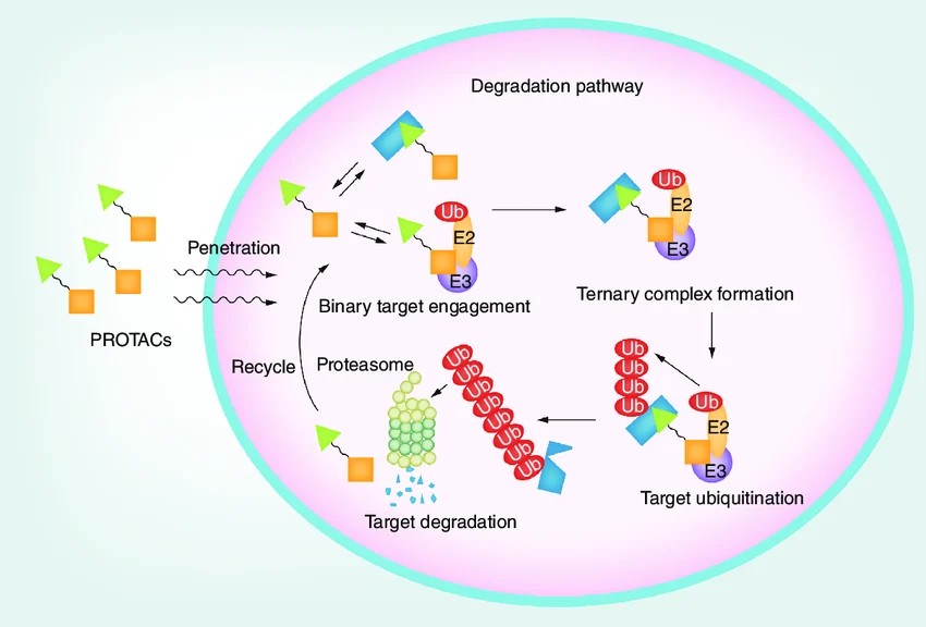 Figure 1. Degradation pathway for proteolysis targeting chimeras. (Liu et al., 2020)