We use cookies to understand how you use our site and to improve the overall user experience. This includes personalizing content and advertising. Read our
Privacy Policy
Profacgen offers high-sensitivity Circular Dichroism (CD) spectroscopy services for quantitative analysis of protein secondary structure, conformational integrity, folding behavior, and thermal stability, using both far-UV and near-UV CD.
CD measures the differential absorption of left- and right-circularly polarized light. In proteins, peptide bond arrangement in regular secondary structures generates characteristic CD bands with distinct positions and intensities, enabling structural determination. This technique is widely used to assess protein folding, the impact of mutations on conformation and stability, and molecular interactions.
Profacgen has established a robust CD platform optimized for both far-UV (secondary structure) and near-UV (tertiary structure) analysis. Our services provide key parameters including stability, folding/unfolding profiles, CD fingerprints, and α-helix/β-sheet content. Near-UV CD (>250 nm) further offers insights into three-dimensional molecular architecture. Our one-stop CD service supports efficient, high-quality structural and interaction analysis for small chiral drugs, metal complexes, and biological macromolecules.
Overview of Circular Dichroism
Circular Dichroism spectroscopy measures the differential absorption of left- and right-handed circularly polarized light by chiral molecules. In proteins and peptides, this differential absorption arises from the asymmetric arrangement of peptide bonds in ordered secondary structures, enabling quantitative structural characterization without crystallization or extensive sample preparation:
Differential absorption of circularly polarized light: The peptide bond chromophore absorbs left- and right-circularly polarized light differently depending on its spatial orientation, generating characteristic CD spectra that encode structural information
Structural characterization of biomolecules: CD provides rapid, non-destructive assessment of secondary structure content, folding state, conformational changes, and thermal stability under native solution conditions
Figure 1. Schematic representation of the Circular Dichroism instrument configuration. (Pignataro, et al., 2020)
CD spectroscopy is particularly powerful because it operates under physiologically relevant conditions, requires minimal sample quantities, and provides real-time monitoring of structural transitions. The technique is applicable across a broad molecular weight range and is compatible with diverse buffer systems, making it an essential tool for protein characterization, formulation development, and comparability assessment.
What Can CD Measure?
Profacgen's CD platform delivers comprehensive quantitative measurements across the critical structural attributes of biomolecular samples:
Secondary Structure Composition: Quantitative estimation of α-helix, β-sheet, β-turn, and random coil content through deconvolution of far-UV CD spectra (190–250 nm), enabling comparison with reference structures and detection of folding anomalies
Protein Folding and Unfolding: Real-time monitoring of conformational transitions induced by chemical denaturants, pH changes, or mechanical stress, providing thermodynamic parameters (ΔG, Cm) and kinetic rate constants for folding mechanism elucidation
Conformational Changes: Detection of subtle structural perturbations caused by mutations, post-translational modifications, formulation components, or storage conditions, enabling quality control and developability assessment
Ligand-Induced Structural Changes: Quantification of binding-induced conformational shifts upon interaction with small molecules, nucleic acids, lipids, or protein partners, supporting mechanism-of-action studies and drug discovery
Thermal Stability: Temperature-dependent CD monitoring to determine melting temperatures (Tm), cooperative unfolding transitions, and thermal stability rankings under formulation-relevant conditions
Figure 2. Representative of far-UV CD spectra and near-UV CD spectra. (Pignataro, et al., 2020)
Our CD Analysis Services
Profacgen provides specialized CD analysis services tailored to diverse sample types and analytical objectives. Each service module is optimized for the specific structural information required and the physicochemical properties of the target biomolecule.
Secondary Structure Determination
Quantitative estimation of protein secondary structure content using far-UV CD spectroscopy with validated deconvolution algorithms.
Proteins: α-helix, β-sheet, β-turn, and random coil quantification for recombinant therapeutic proteins, enzymes, and structural proteins
Peptides: Conformational state assessment (ordered vs. disordered), helical propensity determination, and structure-activity relationship support for peptide drug candidates
Antibodies: Fab and Fc domain structural integrity, complementarity-determining region (CDR) conformation assessment, and biosimilar higher-order structure comparability
Protein Folding Studies
Comprehensive characterization of folding pathways, refolding efficiency, and structural recovery under diverse conditions.
Refolding optimization: Real-time CD monitoring of inclusion body refolding, dilution refolding, and chromatographic refolding to identify conditions maximizing native structure recovery
Structural comparison: Side-by-side CD spectral comparison of wild-type and variant proteins, process intermediates, or stressed samples to detect conformational deviations and guide optimization
Thermal Denaturation Analysis
Temperature-dependent CD monitoring to determine thermal stability parameters and rank formulation or variant stability.
Tm determination: Accurate melting temperature measurement through cooperative unfolding transition monitoring in the far-UV or near-UV range, with van't Hoff or calorimetric analysis
Stability ranking: Comparative thermal stability assessment across antibody candidates, formulation conditions, or manufacturing batches to identify robust, developable molecules
Conformational Change Assessment
Detection and quantification of environmentally or genetically induced structural perturbations.
Mutation studies: CD spectral comparison of wild-type and mutant proteins to assess structural impact of amino acid substitutions, deletions, or insertions on folding and stability
Ligand binding studies: Quantification of binding-induced conformational shifts upon interaction with small molecules, nucleic acids, lipids, or protein partners to support mechanistic understanding
Formulation evaluation: Assessment of buffer, pH, ionic strength, and excipient effects on conformational integrity and stability under storage and stress conditions
Applications
Our CD services support a broad spectrum of applications across biopharmaceutical development, manufacturing, and quality assessment:
Protein Engineering: Secondary structure verification, folding state confirmation, and mutation impact assessment for engineered enzymes, therapeutic proteins, and peptide-based drug candidates
Antibody Characterization: Higher-order structure assessment, Fab/Fc domain integrity verification, CDR conformation monitoring, and aggregation-induced structural change detection for monoclonal, bispecific, and Fc-fusion antibodies
Biosimilar Comparability: Rigorous far-UV and near-UV CD spectral comparison between innovator and candidate products to demonstrate higher-order structure equivalence, a critical quality attribute for regulatory approval
Stability Assessment: Thermal stability profiling, forced degradation structural monitoring, and formulation-induced conformational change detection to support formulation optimization and shelf-life determination
Structure–Function Studies: Correlation of secondary structure content and conformational dynamics with biological activity, binding affinity, and pharmacokinetic properties to guide rational design and optimization
Data Outputs
Profacgen provides structured, decision-ready analytical outputs aligned with your structural characterization and regulatory requirements:
Secondary Structure Estimation: Quantitative α-helix, β-sheet, β-turn, and random coil percentages derived from validated deconvolution algorithms (CONTIN, SELCON, CDSSTR) with goodness-of-fit metrics and confidence intervals
Spectral Profiles: Raw and baseline-corrected far-UV (190–250 nm) and near-UV (250–320 nm) CD spectra with molar ellipticity or mean residue ellipticity units, presented as publication-quality figures with experimental conditions
Thermal Melt Curves: Temperature-dependent ellipticity profiles with fitted transition curves, derivative analysis, and cooperative unfolding assessment for Tm determination and stability ranking
Tm Determination: Accurate melting temperatures with statistical precision, van't Hoff enthalpy calculations, and comparison across samples, conditions, or timepoints for robust stability assessment
Comparative Structural Analysis: Side-by-side spectral overlay, difference spectra, and quantitative similarity metrics (spectral correlation coefficient, root-mean-square deviation) for biosimilar comparability, process change assessment, and stress comparison
High-Sensitivity Spectroscopic Analysis: Optimized CD instrumentation ensures high signal-to-noise ratios in both far- and near-UV ranges, enabling accurate secondary structure quantification and detection of subtle conformational changes affecting activity and stability.
Broad compatibility: Supports proteins, peptides, antibodies, nucleic acids, chiral small molecules, and metal complexes across diverse concentrations and molecular weights.
Customized design: Experimental parameters—wavelength, temperature, buffer, and acquisition settings—are tailored to your molecule and objectives for actionable results.
Expert interpretation: Our team integrates biophysics expertise, CD theory, deconvolution algorithms, and pharmaceutical development experience to translate spectral data into meaningful structural insights and practical recommendations.
A biosimilar development program required rigorous demonstration of higher-order structure equivalence between a candidate monoclonal antibody and the reference innovator product. Regulatory agencies consider higher-order structure a critical quality attribute, and CD spectroscopy—particularly the combination of far-UV and near-UV analysis—is a cornerstone of physicochemical comparability assessment.
Objective:
To execute a comprehensive CD comparability study demonstrating equivalent secondary structure content, tertiary structure fingerprints, and thermal stability profiles between the biosimilar candidate and reference product, supported by appropriate statistical analysis and system suitability documentation.
Approach:
Profacgen conducted far-UV CD (190–250 nm) for secondary structure estimation using three validated deconvolution algorithms (CONTIN, SELCON, CDSSTR) and near-UV CD (250–320 nm) for tertiary structure fingerprint comparison. Multiple independent batches of candidate and reference products were analyzed under identical buffer conditions and instrument parameters. Thermal denaturation experiments (25°C to 90°C) were performed to determine Tm values and compare cooperative unfolding transitions. Spectral similarity was quantified using spectral correlation coefficients and root-mean-square deviation metrics.
Outcome:
The biosimilar candidate demonstrated statistically equivalent secondary structure content (α-helix, β-sheet, random coil percentages within ±3%), near-UV spectral profiles with correlation coefficients >0.98, and Tm values within 0.5°C of the reference product. The comprehensive CD dataset and structured report supported regulatory submission and accelerated the path to clinical development.
Scenario 2: Antibody Formulation Optimization by Thermal Stability Screening
Program Context:
A therapeutic antibody development program required rapid identification of a stable, manufacturable formulation for a high-concentration monoclonal antibody exhibiting marginal thermal stability and visible aggregation under preliminary buffer conditions. Traditional stability studies were too time-consuming to screen the required formulation space.
Objective:
To execute a high-throughput CD-based thermal stability screening campaign evaluating pH, buffer species, ionic strength, and excipient effects on conformational integrity and Tm, enabling data-driven selection of an optimal formulation within an accelerated timeline.
Approach:
Profacgen designed a 48-condition formulation matrix and performed temperature-dependent far-UV CD monitoring (25°C to 85°C) for each condition. Thermal melt curves were fitted to determine Tm values, and secondary structure content at 25°C was quantified to assess native state integrity. Selected lead formulations were subjected to accelerated stress studies (40°C, agitation, freeze-thaw) with longitudinal CD monitoring to confirm long-term conformational stability. Near-UV CD was used to assess tertiary structure preservation under optimal conditions.
Outcome:
The screening identified a histidine-sucrose formulation at pH 6.0 that increased Tm by 8°C relative to the initial buffer, maintained native secondary structure content (>95% ordered structure), and showed no detectable conformational change under all stress conditions. The CD-based approach compressed the formulation timeline from months to weeks and conserved development material by 75% compared to traditional stability studies.
Q: What is the difference between far-UV and near-UV CD?
A: Far-UV CD (190–250 nm) primarily probes the peptide bond chromophore and is used for secondary structure determination (α-helix, β-sheet, turns, random coil). Near-UV CD (250–320 nm) probes the aromatic amino acid side chains (Trp, Tyr, Phe) and disulfide bonds, providing information about tertiary structure, asymmetric environment, and protein folding state. Profacgen offers both far-UV and near-UV CD analysis for comprehensive structural characterization.
Q: What sample concentration and volume are required for CD analysis?
A: For far-UV CD, protein concentrations of 0.1–0.5 mg/mL in 10–20 µL are typically sufficient, depending on molecular weight and pathlength. Near-UV CD requires higher concentrations (0.5–2.0 mg/mL) due to weaker aromatic chromophore signals. We recommend samples in low-absorbance buffers (avoid high chloride concentrations) and can advise on optimal preparation conditions for your specific molecule.
Q: How accurate is CD-based secondary structure estimation?
A: When performed with high-quality spectra (190–250 nm, good signal-to-noise) and validated deconvolution algorithms, CD secondary structure estimation typically achieves correlations of 0.90–0.95 with X-ray crystallography and NMR reference structures. Accuracy depends on spectral quality, wavelength range, algorithm selection, and reference dataset compatibility. Profacgen uses multiple algorithms (CONTIN, SELCON, CDSSTR) and reports consensus estimates with confidence metrics.
Q: Can CD detect subtle conformational changes caused by mutations or formulation?
A: Yes. CD is highly sensitive to changes in backbone geometry and aromatic environment. Even single amino acid substitutions can alter CD spectra if they affect secondary structure or tertiary packing. Formulation-induced changes (pH, ionic strength, excipients) are readily detectable through spectral comparison, thermal stability shifts, and secondary structure content changes. Profacgen employs difference spectra and statistical metrics to quantify subtle but significant conformational perturbations.
Q: What is the relationship between Tm and protein stability?
A: Tm (melting temperature) is the temperature at which 50% of the protein population is unfolded under equilibrium conditions. Higher Tm values generally indicate greater thermal stability and resistance to unfolding. Tm is influenced by intrinsic protein sequence, post-translational modifications, formulation conditions, and ligand binding. In developability assessment, Tm ranking helps identify stable candidates and robust formulations. However, Tm should be interpreted alongside kinetic stability and aggregation data for comprehensive stability evaluation.
Q: How does CD compare to other structural techniques like X-ray crystallography or NMR?
A: CD provides rapid, low-material, solution-phase structural information without crystallization requirements, making it ideal for screening, stability monitoring, and comparability assessment. However, CD provides lower-resolution structural information (secondary structure content rather than atomic coordinates) compared to X-ray crystallography or NMR. CD is best used as a complementary technique—for rapid assessment, quality control, and monitoring—while high-resolution techniques provide detailed atomic structures. Profacgen integrates CD data with other biophysical techniques for comprehensive characterization.
References:
Pignataro MF, Herrera MG, Dodero VI. Evaluation of peptide/protein self-assembly and aggregation by spectroscopic methods. Molecules. 2020;25(20):4854. doi:10.3390/molecules25204854
Online Inquiry
Fill out this form and one of our experts will respond to you within one business day.
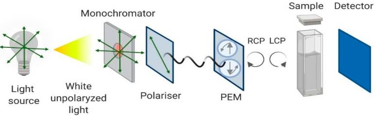 Figure 1. Schematic representation of the Circular Dichroism instrument configuration. (Pignataro, et al., 2020)
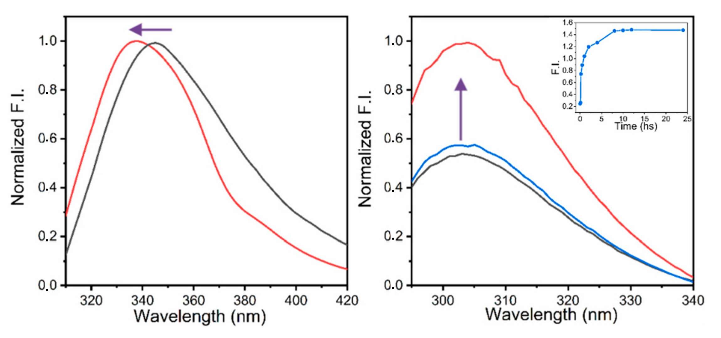 Figure 2. Representative of far-UV CD spectra and near-UV CD spectra. (Pignataro, et al., 2020)