
Understanding the precise amino acid sequence that mediates a protein-protein interaction is essential for rational drug design, epitope mapping, and mechanistic studies. Profacgen's protein binding site mapping service uses surface plasmon resonance (SPR) spectroscopy combined with overlapping peptide libraries to pinpoint the exact binding region on your target protein. This label-free, real-time approach delivers quantitative binding data with peptide-level resolution, enabling structure-guided optimization of therapeutic candidates.
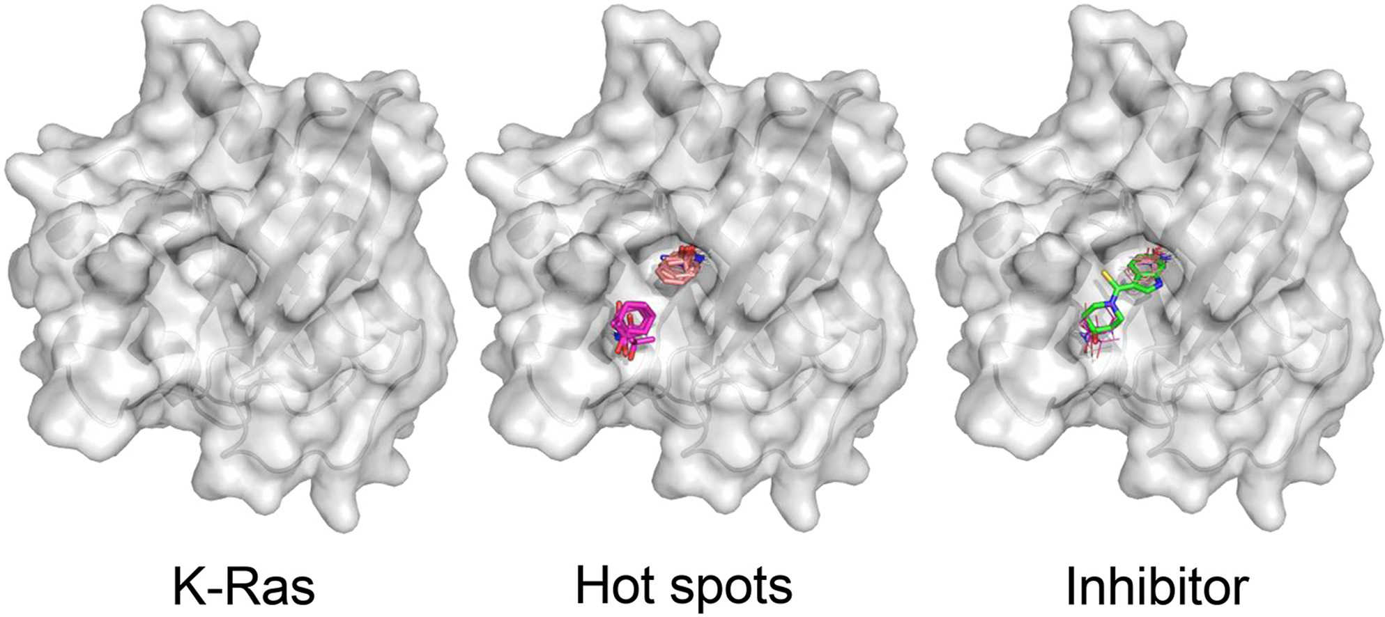 Figure 1. Exmaple of protein binding site mapping. (Wakefield et al., 2022)
Surface plasmon resonance (SPR) is an optical detection method that measures the refractive index change near a gold sensor surface when molecules bind. In a typical SPR experiment, one binding partner (ligand) is immobilized on the sensor chip while the other (analyte) flows through the microfluidic channel. Binding is detected in real time as a change in the SPR angle, producing a sensorgram that reveals both binding kinetics (association and dissociation rates) and affinity (equilibrium dissociation constant, KD).
For binding site mapping, the target protein sequence is tiled with overlapping peptides (typically 15- or 20-mers with 5-10 residue overlaps) that collectively cover the entire protein. The peptide library is immobilized on the SPR sensor surface as an array of spatially defined spots. The analyte protein is injected across the peptide array, and binding is detected at each spot in parallel. Peptides that generate a positive SPR signal correspond to the regions of the target protein where binding occurs.
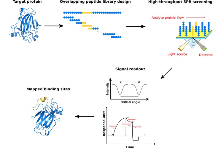
Profacgen's high-throughput SPR workflow enables rapid screening of hundreds of overlapping peptides in a single experiment, with positive and negative controls and replicate spots ensuring data confidence. The resulting binding map provides the molecular basis for interaction mechanism studies and targeted therapeutic design.
Profacgen provides comprehensive binding site mapping services for research and drug discovery applications. Our offerings include:
| Service Component | Description |
|---|---|
| Standard Binding Site Mapping | SPR screening of 15-mer overlapping peptide library covering full target sequence. Deliverables: binding heatmap, positive peptide list, binding kinetics, comprehensive report. |
| High-Resolution Mapping | Finer peptide tiling (10-mer overlaps) for precise epitope delineation. Deliverables: refined binding region, kinetic analysis, alanine scan recommendations. |
| Alanine Scanning Mutagenesis | Systematic substitution of binding region residues to identify critical contacts. Deliverables: key residue map, interaction hot spot identification, energy contribution ranking. |
| Competition Epitope Binning | Determine whether multiple antibodies bind overlapping or distinct epitopes. Deliverables: epitope binning matrix, pairwise competition classification. |

To complement your protein interaction analysis, explore our comprehensive portfolio of related screening and profiling services.
Background:
A vaccine developer needed to map the binding epitope of a potent neutralizing antibody against the SARS-CoV-2 spike protein receptor-binding domain (RBD). Precise epitope definition was required to support vaccine immunogen design and intellectual property claims.
Our Solution:
Using our SPR-based binding site mapping platform, we designed a library of 15-mer peptides tiling the entire RBD sequence (319 amino acids, 61 peptides). The peptide library was immobilized on a CM5 sensor chip and screened against the antibody at multiple concentrations. Positive and negative controls were included for data validation.
Final Results:
SPR screening identified three adjacent peptides generating positive binding signals, defining a continuous epitope spanning residues 438-450. Alanine scanning of this region revealed two critical contact residues (F456 and Y473) whose mutation abolished binding. The epitope data informed the client's vaccine immunogen design and supported intellectual property filing.
Background:
A structural biology group required precise definition of the binding interface between an interleukin cytokine and its alpha receptor subunit to guide the design of receptor antagonists. The interaction was known to be essential for inflammatory signaling but the precise contact residues were undefined.
Our Solution:
We generated overlapping peptide libraries for both proteins and performed reciprocal SPR binding site mapping. The cytokine library (156 amino acids, 29 peptides) and receptor library (342 amino acids, 66 peptides) were screened against each other to identify complementary binding patches.
Final Results:
The screen identified a 25-residue binding patch on the cytokine and a complementary 18-residue region on the receptor. Competition experiments with a known antagonist confirmed overlap with the mapped interface. The data were used to build a structural model that predicted antagonist binding poses with high accuracy, accelerating the client's lead optimization program by approximately six months.
Get a Quote for your protein site mapping project
References:
Fill out this form and one of our experts will respond to you within one business day.