We use cookies to understand how you use our site and to improve the overall user experience. This includes personalizing content and advertising. Read our
Privacy Policy
At Profacgen, our protein conjugation service on beads provides high-quality, reliable coupling of proteins, antibodies, and other biomolecules to superparamagnetic and non-magnetic microspheres for applications in affinity purification, immunoprecipitation, immunoassays, cell isolation, and nucleic acid extraction. Magnetic bead technology has become the platform of choice for modern biomolecular separation due to its unique combination of rapid magnetic responsiveness, homogeneous suspension behavior, and straightforward integration with automated liquid handling systems.
Profacgen supplies several types of beads with distinct surface chemistries and functional properties. Each bead type is optimized for specific applications, from the high-affinity capture of biotinylated molecules using streptavidin-coated beads to the versatile covalent coupling enabled by tosyl-activated or epoxy-functionalized surfaces. Our conjugation protocols are validated to ensure high coupling efficiency, minimal protein denaturation, and consistent batch-to-batch performance, with comprehensive quality control including coupling capacity determination, leaching tests, and functional validation.
Background: Magnetic Bead Technology for Biomolecular Separation
Superparamagnetic beads comprise iron oxide nanocrystals (Fe3O4 or maghemite, γ-Fe2O3) embedded in a polymer matrix (polystyrene, polyacrylate, or agarose). They magnetize rapidly under an external field for quick separation yet retain no residual magnetism upon field removal, ensuring easy resuspension without aggregation.
Bead size influences performance: smaller beads (1–2 µm) offer higher surface area and faster binding kinetics but require stronger magnetic fields; larger beads (2.8–4.5 µm) enable faster separation, ideal for automated workflows. Uniform size distribution ensures consistent magnetic response and batch-to-batch reproducibility.
Surface chemistry dictates coupling options and non-specific binding. Hydrophilic surfaces (agarose, hydroxyl-modified silica) minimize protein adsorption, favoring purification applications. Hydrophobic surfaces (polystyrene) offer higher binding capacity but increased non-specific binding, better suited for assay applications where capture specificity dominates. Profacgen selects optimal bead type and surface chemistry based on project-specific needs.
Figure 1. An overview of the magnetic bead-based separation (MBS) method. (York et al., 2023)
Our Bead Product Portfolio
Profacgen provides the following bead types for protein conjugation:
Streptavidin MagPoly Beads
High-capacity streptavidin-coated magnetic beads for capture of biotinylated proteins, antibodies, nucleic acids, and other biomolecules via the ultra-high-affinity streptavidin-biotin interaction.
Bead size: 2.8 μm (optimal for rapid magnetic separation)
Streptavidin density: 8–12 pmol/mg beads
Biotin-binding capacity: 800–1,200 pmol/mg
Working pH: 4–9; elution: harsh conditions or competitive biotin
Tosyl-activated beads for covalent coupling of proteins and peptides through amine or thiol groups, providing stable, leach-resistant attachment suitable for reusable applications.
Bead size: 2.8 μm
Surface: tosyl groups reactive toward NH2 and SH at pH 7–9
Coupling capacity: 10–30 μg protein/mg beads
Linkage: stable secondary amine or thioether bond
Applications: antibody coupling for IP, enzyme immobilization, assay development
Ni-NTA MagPoly Beads
Nickel-charged nitrilotriacetic acid beads for affinity capture of His-tagged recombinant proteins with high specificity and convenient imidazole elution.
Applications: protein purification, pull-down assays, enzymatic assays
Protein A/G MagPoly Beads
Recombinant Protein A or Protein G coated beads for oriented immobilization of antibodies via Fc-region binding, maximizing antigen-binding accessibility.
Bead size: 2.8 μm
Binding capacity: 25–40 mg IgG/mL settled beads
Elution: pH 2–3 (glycine-HCl) or milder with Protein G
Crosslinking option available for permanent antibody attachment
Silica-coated magnetic beads for nucleic acid extraction and purification, binding DNA/RNA through chaotropic salt-mediated interactions.
Bead size: 200–800 nm (optimal for DNA/RNA binding surface area)
Surface: silicon hydroxyl groups for nucleic acid adsorption
Binding: hydrogen bonding and electrostatic interactions in high-salt/low-pH conditions
Elution: low-salt buffer or water
Applications: plasmid extraction, PCR cleanup, gel recovery, viral DNA/RNA isolation
Agarose Matrix Microspheres
Magnetic agarose beads combining high porosity with low non-specific binding, ideal for affinity purification of sensitive proteins.
Bead size: 30–100 μm (porous agarose matrix)
Surface: abundant hydroxyl groups for CNBr or NHS activation
High binding capacity due to internal surface area
Low non-specific protein adsorption
Applications: protein purification, affinity chromatography, enzyme immobilization
Applications
Affinity purification: Capture His-tagged, biotinylated, or antibody-targeted proteins from crude lysates, serum, or culture supernatant with high purity and yield
Immunoprecipitation (IP) and co-IP: Rapid isolation of target proteins and interaction partners from cell lysates using antibody-conjugated beads; amenable to downstream Western blot or mass spectrometry analysis
Cell isolation and sorting: Positive or negative selection of cell populations (CD4+ T cells, CD14+ monocytes, circulating tumor cells) using antibody-coated beads; compatible with clinical-grade cell therapy manufacturing
Nucleic acid extraction: High-throughput, automatable DNA/RNA isolation from blood, tissue, cell culture, and environmental samples using MagSilica beads
Immunoassay development: Antibody-conjugated beads as capture reagents in luminescent oxygen channeling (LOCI), electrochemiluminescence (ECL), and bead-based flow cytometry assays
Phage display biopanning: Enrichment of phage clones binding to immobilized target proteins with high efficiency and low background
Representative Case Studies
Case Study 1: Streptavidin Bead-Based mRNA Capture for Single-Cell Sequencing
Background:
A single-cell genomics company required a streptavidin bead system for capturing polyadenylated mRNA using biotinylated oligo(dT) primers in their high-throughput single-cell RNA-seq workflow. The beads needed to show >95% capture efficiency, minimal rRNA contamination, and compatibility with automated liquid handlers.
Approach:
Profacgen evaluated three streptavidin bead densities (low, medium, high) for mRNA capture efficiency from 10–1,000 cell equivalents. Optimal biotinylated oligo(dT) loading was determined by titration. Beads were processed on an automated platform with magnetic separation modules.
Outcome:
High-density streptavidin beads (1,100 pmol biotin-binding/mg) achieved 97.3% mRNA capture efficiency from 100 pg total RNA input. rRNA contamination was <2% (verified by Bioanalyzer). The automated workflow processed 384 samples in 4 hours with CV <5% across replicates. The system was validated against the company's previous column-based method, showing equivalent gene detection sensitivity (median 4,500 genes/cell) with 3-fold higher throughput. The bead-based workflow is now the standard for the company's commercial single-cell sequencing kit.
Case Study 2: Enzyme-Coupled Tosyl Beads for a Diagnostic Metabolite Test
Background:
A point-of-care diagnostic company needed to immobilize glucose oxidase (GOx) and horseradish peroxidase (HRP) on tosyl-activated magnetic beads for a dual-enzyme cascade reaction in their glucose test strip. The beads needed to maintain >90% enzyme activity after 18 months of dry storage at room temperature.
Approach:
Profacgen co-immobilized GOx and HRP on 2.8 μm tosyl beads at a 2:1 molar ratio using optimized coupling conditions (pH 7.4, 18 h, 4°C). After coupling, free enzyme was removed by magnetic washing and the beads were lyophilized with trehalose as a stabilizer.
Outcome:
Enzyme coupling efficiency was 85% for GOx and 78% for HRP. The co-immobilized beads retained 94% of initial dual-enzyme activity after 18 months of storage at 25°C in dry form—exceeding the target specification. The bead-based test showed a linear range of 0.5–30 mM glucose with R2 = 0.999, matching the performance of the company's previous liquid enzyme formulation while eliminating cold-chain storage requirements. The test received CE-IVD marking and is deployed in over 500 clinics.
Q: How do I choose the right bead type for my application?
A: Selection depends on your target molecule and application. For biotinylated proteins or nucleic acids: streptavidin beads. For His-tagged proteins: Ni-NTA beads. For antibody purification or IP: Protein A/G beads. For covalent, irreversible protein coupling: tosyl-activated or epoxy beads. For nucleic acid extraction: MagSilica beads. For sensitive protein purification: agarose matrix beads. We provide consultation to match the optimal bead type, surface chemistry, and conjugation protocol to your specific requirements, and can supply small trial quantities for evaluation.
Q: What bead sizes are available and how does size affect performance?
A: We offer beads from 200 nm to 100 μm. Sub-micron beads (200–800 nm, e.g., MagSilica) offer the highest surface area-to-volume ratio for maximum binding capacity and are ideal for nucleic acid extraction. Micron-sized beads (1–4.5 μm) provide the best balance of binding capacity and rapid magnetic separation, suitable for most protein and cell applications. Large porous beads (30–100 μm, e.g., agarose) provide very high binding capacity due to internal porosity but require longer separation times and are best for batch-mode purification. We recommend 2.8 μm beads as the default for protein applications and 200–500 nm for nucleic acid work.
Q: How do you validate protein coupling efficiency and stability?
A: We use a multi-parameter validation approach: (1) coupling efficiency determined by comparing protein concentration in supernatant before and after coupling (BCA or A280); (2) binding capacity measured by titrating the target analyte and determining the saturation point; (3) leaching test by incubating coupled beads in working buffer and measuring released protein over time; (4) functional validation using a relevant assay (e.g., antigen binding for antibody beads, enzymatic activity for enzyme beads); and (5) storage stability evaluated by monitoring activity over 12–24 months under recommended storage conditions. A comprehensive QC report is provided with each batch.
Q: Can you provide custom bead functionalization?
A: Yes. We offer custom surface functionalization including: introduction of specific reactive groups (azide, alkyne, tetrazine, hydrazide, aldehyde); coating with specific affinity ligands (custom antibodies, lectins, aptamers); fluorescent dye doping for multiplexed assays; size customization within manufacturing constraints; and GMP-grade bead production with full traceability and documentation. Custom projects typically require 4–8 weeks from specification to delivery. Please contact us to discuss your custom bead requirements.
Q: Are your beads compatible with automated platforms?
A: Yes. Our magnetic beads are compatible with all major automated liquid handling platforms including Hamilton, Tecan, Beckman Coulter Biomek, Thermo KingFisher, and PerkinElmer Zephyr. We provide validated protocols for common applications (nucleic acid extraction, IP, bead-based ELISA) on these platforms. The 2.8 μm size provides optimal magnetic separation speed for most automated magnetic separators. We can also develop and validate custom automated workflows for high-throughput applications.
References:
York A, Huynh E, Mbodj S, et al. Magnetic bead-based separation of Pneumococcal serotypes. Cell Reports Methods. 2023;3(2):100410. doi:10.1016/j.crmeth.2023.100410
Online Inquiry
Fill out this form and one of our experts will respond to you within one business day.

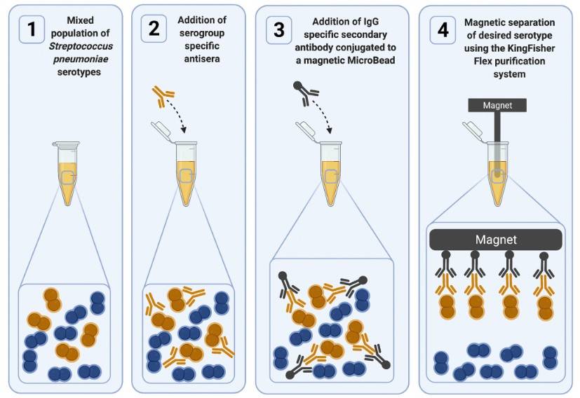 Figure 1. An overview of the magnetic bead-based separation (MBS) method. (York et al., 2023)