We use cookies to understand how you use our site and to improve the overall user experience. This includes personalizing content and advertising. Read our
Privacy Policy
Bioluminescence resonance energy transfer (BRET) is a highly sensitive, proximity-based biophysical technique that enables real-time monitoring of molecular interactions within living cells. The method exploits non-radiative dipole-dipole energy transfer from a bioluminescent donor enzyme—typically a luciferase—to a fluorescent acceptor molecule. When the donor and acceptor are fused to proteins of interest and brought into close proximity (typically <10 nm), energy transfer occurs, producing a quantifiable emission signal at the acceptor wavelength. This ratiometric readout directly reflects the spatial proximity and interaction dynamics of the tagged partners in their native cellular environment. The emergence of engineered compact luciferases (small-form luciferase) and optimized acceptor fluorophores has revolutionized BRET detection through enhanced brightness, spectral separation, and signal stability, enabling robust interrogation of protein-protein interactions, protein-ligand binding, and receptor conformational changes with unprecedented temporal and spatial resolution. Profacgen provides comprehensive BRET assay services leveraging conventional BRET, BRET1, BRET2, eBRET, and next-generation enhanced BRET platforms, integrated with advanced molecular biology and live-cell imaging capabilities to support drug discovery, GPCR pharmacology, and mechanistic cell biology research.
Principle and Instrumentation
BRET is grounded in Förster resonance energy transfer (FRET) physics, with the critical distinction that excitation energy originates from an enzymatic chemiluminescent reaction rather than external illumination. This fundamental difference eliminates phototoxicity, autofluorescence, and photobleaching—limitations that constrain conventional FRET in live-cell and long-duration experiments.
Figure 1. The principle of bioluminescence resonance energy transfer (BRET) for monitoring biological proximity and setup for BRET microscopy. (Kobayashi et al., 2019)
Energy Transfer Mechanism
The BRET signal arises from the following sequence:
Substrate Oxidation: The luciferase donor catalyzes oxidation of a specific substrate (coelenterazine for conventional BRET; furimazine for enhanced BRET), producing an excited-state oxyluciferin intermediate
Photon Emission and Dipole Coupling: When the acceptor fluorophore is within the Förster radius (R0), non-radiative dipole-dipole coupling transfers excitation energy from donor to acceptor with efficiency proportional to the inverse sixth power of intermolecular distance
Ratiometric Detection: The BRET ratio is calculated as acceptor emission intensity divided by donor emission intensity, normalizing for expression level variations and providing a robust, internally controlled proximity metric
BRET Platform Configurations
Profacgen operates multiple BRET variants optimized for distinct experimental requirements:
Platform
Donor
Acceptor
Substrate
Key Features
Conventional BRET
Renilla luciferase (Rluc, 37 kDa)
YFP or GFP variants
Coelenterazine h or 400a
Established methodology; moderate brightness; suitable for strong interactions
BRET1
Renilla luciferase
YFP (eYFP, Venus)
Coelenterazine h
Maximal spectral overlap for highest signal amplitude; broader emission overlap requires careful background subtraction
BRET2
Renilla luciferase
GFP2
Coelenterazine 400a
Enhanced spectral separation (415 nm donor, 515 nm acceptor); reduced background but lower absolute signal
eBRET
Enhanced Rluc variants
Red-shifted acceptors (mOrange, mCherry)
Coelenterazine derivatives
Extended spectral window; reduced compound interference; improved tissue penetration for in vivo applications
Enhanced BRET
small-form luciferase (19 kDa, 150× brighter than Rluc)
self-labeling protein tag or SNAP-tag fluorophores
Furimazine
Optimal spectral overlap with minimal background; increased signal-to-noise; compatible with extracellular luciferase inhibitors for signal tuning
The compact size of small-form luciferase (19 kDa versus 37 kDa for Rluc) minimizes steric disruption of fusion protein folding and trafficking, while its exceptional brightness enables detection of low-abundance interactions and transient complexes that are invisible to conventional BRET systems.
Applications
BRET technology has become indispensable across multiple domains of molecular and cellular research, particularly where physiological relevance and dynamic monitoring are paramount.
Live-Cell Protein-Protein Interaction Mapping
Real-time detection of binary and multimeric protein complexes in their native subcellular compartments. BRET enables tracking of interaction kinetics, dissociation rates, and complex stoichiometry without cell lysis or fixation artifacts. Applications include kinase-substrate recognition, transcription factor dimerization, and scaffold protein assembly dynamics.
GPCR Pharmacology and Signal Transduction
Quantitative analysis of GPCR oligomerization, ligand-induced conformational changes, G protein coupling specificity, and β-arrestin recruitment. BRET biosensors report receptor activation state through intramolecular conformational BRET (conBRET) or intermolecular coupling BRET, supporting drug candidate profiling and allosteric modulator characterization.
Protein-Ligand and Protein-Drug Binding
Competitive and direct binding assays for small molecule inhibitors, peptide ligands, and therapeutic antibodies. Enhanced BRET target engagement assays use tracer ligands to quantify compound occupancy at endogenous protein targets in live cells, bridging biochemical affinity and cellular pharmacology.
Receptor Trafficking and Internalization
Monitoring of agonist-induced receptor endocytosis, recycling, and degradation through BRET between surface-localized and intracellular compartments. Real-time trafficking profiles distinguish biased agonists with distinct desensitization and resensitization kinetics.
Epigenetic and Chromatin Proximity Analysis
BRET-based proximity labeling for mapping protein associations with chromatin modifications, histone variants, and DNA regulatory elements in living nuclei. Complementary to ChIP-seq by preserving transient interactions lost during crosslinking and extraction.
In Vivo and Ex Vivo Imaging
Deep-tissue BRET imaging using red-shifted eBRET and near-infrared luciferase variants for preclinical pharmacodynamic studies, tumor microenvironment interaction mapping, and host-pathogen proximity detection in animal models.
Technical Advantages
Native Cellular Environment: Measurements performed in intact, living cells preserve post-translational modifications, native folding, and correct subcellular localization unavailable in cell-free systems
Exceptionally Low Background: Absence of excitation illumination eliminates cellular autofluorescence, scatter, and phototoxicity; bioluminescent emission provides near-zero background for high signal-to-noise discrimination
Real-Time Kinetic Resolution: Continuous monitoring of association and dissociation dynamics with temporal resolution from seconds to hours, capturing transient intermediates and equilibrium shifts upon ligand addition or washout
Ratiometric Internal Control: BRET ratio normalization corrects for cell number, transfection efficiency, and expression level variations, enabling quantitative comparison across experiments and laboratories
Minimal Steric Perturbation: The small-form luciferase's 19 kDa size and engineered fusion compatibility reduce disruption of target protein function compared to larger reporter enzymes or fluorescent protein fusions
Pharmacological Tunability: Extracellular luciferase inhibitors (e.g., luciferase inhibitor, PTC124 derivatives) enable acute signal modulation, background suppression, and temporal control of BRET reporter activity
Service Procedure
Profacgen delivers end-to-end BRET services encompassing molecular construct design, cell line engineering, assay optimization, high-throughput screening, and mechanistic data interpretation. Our workflow ensures robust, reproducible proximity measurements aligned with project-specific scientific objectives.
Construct Design and Molecular Cloning: Fusion of small-form luciferase, Rluc, or self-labeling protein tag/SNAP-tag moieties to N-terminus, C-terminus, or internal loops of target proteins based on structural topology and functional domain mapping; linker optimization to maximize energy transfer while preserving biological activity
Cell Line Generation and Validation: Stable or transient transfection into physiologically relevant cell backgrounds (HEK293, CHO, U2OS, primary cells); monoclonal selection for stable lines with uniform expression; Western blot and flow cytometry confirmation of fusion protein integrity and localization
Assay Development and Optimization: Substrate concentration titration, emission filter selection, integration time optimization, and Z'-factor determination for screening readiness; donor-acceptor expression ratio balancing to maximize dynamic range
High-Throughput Screening Execution: Automated liquid handling and plate-based BRET detection for compound library screening, mutagenesis profiling, or interaction mapping; single-concentration primary screens followed by dose-response confirmation
Pharmacological Characterization: Competitive binding curves, kinetic association/dissociation profiles, and allosteric modulation assessment using tracer displacement and conformational biosensor formats
Data Analysis and Mechanistic Interpretation: BRET ratio calculation, saturation binding modeling, kinetic fitting, and structure-activity relationship integration with downstream crystallography or molecular dynamics studies
Case 1: Enhanced BRET-Based GPCR Heterodimerization Screen for Pain Therapeutic Discovery
Background:
A pharmaceutical company sought to identify small molecule modulators that selectively disrupt pathological heterodimerization between the μ-opioid receptor (MOR) and the chemokine receptor CXCR4, a complex implicated in opioid tolerance and hyperalgesia. Conventional co-immunoprecipitation and FRET approaches failed to detect this transient interaction with sufficient sensitivity for compound screening.
Our Solution:
Profacgen engineered an enhanced BRET system with MOR-small luciferase and CXCR4-self-labeling tag fusions, validated for correct plasma membrane trafficking and ligand binding functionality. We optimized the donor-acceptor expression ratio and furimazine concentration to achieve a robust BRET signal responsive to CXCL12-induced heterodimerization. A 10,000-compound library was screened in 384-well format, with hits confirmed by orthogonal BRET saturation and kinetic dissociation assays.
Final Results:
The enhanced BRET screen identified 23 compounds that reduced MOR-CXCR4 BRET by >50% without affecting MOR homodimerization controls. Lead optimization of the top chemotype yielded a candidate with 180 nM IC50 in the heterodimer disruption assay and no detectable MOR agonism or antagonism. The compound advanced to in vivo pain model evaluation, demonstrating reduced tolerance liability compared to morphine in a chronic constriction injury model.
Case 2: Intracellular Target Engagement Profiling by Enhanced BRET Tracer Displacement
Background:
A kinase inhibitor development program required cellular target engagement confirmation for a series of CDK2 inhibitors with sub-nanomolar biochemical potency. Traditional cellular pharmacology assays (phospho-Rb, cell cycle arrest) showed poor correlation with biochemical affinity, suggesting compound permeability or intracellular target accessibility issues that biochemical assays could not resolve.
Our Solution:
Profacgen established an enhanced BRET target engagement assay by fusing small-form luciferase to the N-terminus of CDK2 and introducing a cell-permeable fluorescent tracer ligand that binds the ATP-binding site. Inhibitor displacement of the tracer was quantified as a reduction in BRET signal, directly reporting intracellular target occupancy. We profiled 45 compounds across a 10-point concentration range in live HEK293 cells, with parallel biochemical competition and cellular permeability measurements.
Final Results:
Enhanced BRET target engagement revealed that 12 of 45 compounds with <1 nM biochemical Ki showed >100-fold weaker cellular target engagement, tracing the discrepancy to efflux transporter susceptibility (MDR1 substrate identification) or lysosomal trapping. Two compounds with balanced biochemical potency and cellular engagement were prioritized, and their enhanced BRET-derived cellular IC50 values accurately predicted in vivo tumor growth inhibition in a CDK2-dependent xenograft model. The target engagement data supported the client's target product profile and investor due diligence.
Q: What is the difference between BRET and FRET for protein interaction analysis?
A: Both BRET and FRET detect molecular proximity through resonance energy transfer, but they differ fundamentally in excitation source. FRET requires external illumination to excite the donor fluorophore, introducing phototoxicity, autofluorescence background, and photobleaching that limit live-cell applications. BRET uses enzymatic bioluminescence as the excitation source, eliminating these artifacts and providing superior signal-to-noise for long-duration and sensitive live-cell measurements. BRET is also technically simpler, requiring no specialized illumination optics or alignment. However, FRET offers greater flexibility in donor-acceptor spectral pairing and is more readily combined with fluorescence microscopy for spatial resolution.
Q: Can BRET detect transient or weak protein interactions?
A: Yes, particularly with enhanced BRET technology. The exceptional brightness of small-form luciferase (approximately 150-fold brighter than conventional Renilla luciferase) enables detection of interactions with micromolar Kd values and brief residence times that are invisible to standard BRET or co-immunoprecipitation. Real-time kinetic BRET measurements can capture association and dissociation events on sub-second to minute timescales. For extremely transient interactions, we can employ conformational BRET (intramolecular BRET within a single polypeptide) or stabilized scaffolds to increase effective local concentration.
Q: How does enhanced BRET achieve lower background than conventional BRET?
A: Enhanced BRET achieves background reduction through three mechanisms: (1) optimal spectral overlap between small-form luciferase emission (peak 460 nm) and the self-labeling protein tag or SNAP-tag acceptor excitation spectrum, maximizing energy transfer efficiency while minimizing direct acceptor excitation; (2) the use of cell-permeable luciferase inhibitors that suppress non-specific donor emission without affecting the BRET signal from properly assembled complexes; and (3) the small size of small-form luciferase (19 kDa), which reduces non-specific aggregation and mislocalization that can generate false-positive proximity signals in conventional BRET systems.
Q: What controls are essential to validate a BRET interaction?
A: Rigorous BRET validation requires multiple control experiments: (1) donor-only and acceptor-only transfections to establish baseline emission spectra and exclude spectral bleed-through; (2) negative controls using non-interacting protein pairs or point mutations known to disrupt the interaction; (3) positive controls with well-characterized interacting partners; (4) BRET saturation analysis to demonstrate signal saturation at high acceptor-donor ratios, confirming specific interaction rather than random collision; and (5) pharmacological validation using known inhibitors or competing ligands to demonstrate signal reversibility. Profacgen implements all standard controls and can design custom validation strategies for novel interaction systems.
Q: Can BRET be used for endogenous protein interaction mapping without overexpression?
A: Direct BRET requires genetic fusion of donor and acceptor moieties, necessitating either overexpression or targeted genome engineering. For endogenous interaction mapping, Profacgen employs site-specific endogenous tagging of luminescent or self-labeling reporter proteins at native genomic loci, preserving physiological expression levels and regulatory contexts. Alternatively, we offer BRET-based proximity labeling approaches where a resonance energy transfer pair is recruited to endogenous targets through affinity binders or antibody-directed localization, enabling interaction mapping without permanent genetic modification. These advanced formats require additional development but provide highly physiologically relevant interaction data.
Q: What is the typical project timeline for BRET assay development?
A: Standard enhanced BRET assay development from construct design to validated screening-ready format typically requires 6–8 weeks: 2 weeks for construct generation and sequence verification, 2 weeks for stable cell line establishment and expression validation, 2 weeks for assay optimization and pharmacological benchmarking, and 1–2 weeks for pilot screening and protocol finalization. Expedited timelines (4–5 weeks) are achievable using transient transfection formats for exploratory projects. Complex GPCR signaling assays with multiple BRET biosensors (G protein coupling, β-arrestin recruitment, and receptor internalization) may extend to 10–12 weeks for full panel development and cross-assay validation.
Kobayashi H, Picard LP, Schönegge AM, Bouvier M. Bioluminescence resonance energy transfer–based imaging of protein–protein interactions in living cells. Nat Protoc. 2019;14(4):1084-1107. doi:10.1038/s41596-019-0129-7
Online Inquiry
Fill out this form and one of our experts will respond to you within one business day.
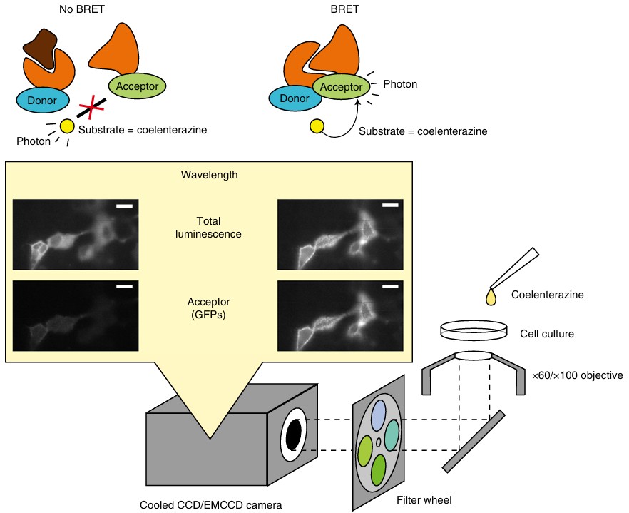 Figure 1. The principle of bioluminescence resonance energy transfer (BRET) for monitoring biological proximity and setup for BRET microscopy. (Kobayashi et al., 2019)