We use cookies to understand how you use our site and to improve the overall user experience. This includes personalizing content and advertising. Read our
Privacy Policy
< 1.0 EU per 1 microgram of protein (determined by LAL method)
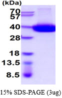
Purity
> 90% by SDS - PAGE
Storage
Can be stored at +4centigrade short term (1-2 weeks). For long term storage, aliquot and store at -20Centigrade or -70Centigrade. Avoid repeated freezing and thawing cycles.
Concentration
1mg/ml (determined by Absorbance at 280nm)
Warning
For research use only!
Background
The protein encoded by this gene is a non-lineage-restricted, type II transmembrane glycoprotein that synthesizes and hydrolyzes cyclic adenosine 5'-diphosphate-ribose, an intracellular calcium ion mobilizing messenger. The release of soluble protein and the ability of membrane-bound protein to become internalized indicate both extracellular and intracellular functions for the protein. This protein has an N-terminal cytoplasmic tail, a single membrane-spanning domain, and a C-terminal extracellular region with four N-glycosylation sites. Crystal structure analysis demonstrates that the functional molecule is a dimer, with the central portion containing the catalytic site. It is used as a prognostic marker for patients with chronic lymphocytic leukemia. Alternative splicing results in multiple transcript variants.
References
Funaro A., et al. (1997) Tissue Antigens. 49:7-15. Lischke., et al. (2013) Infect Immun. 81:4091-4099.
Tag
His tag
Species
Human
Source
Insect cells
BACKGROUND
Background
The protein encoded by this gene is a non-lineage-restricted, type II transmembrane glycoprotein that synthesizes and hydrolyzes cyclic adenosine 5'-diphosphate-ribose, an intracellular calcium ion mobilizing messenger. The release of soluble protein and the ability of membrane-bound protein to become internalized indicate both extracellular and intracellular functions for the protein. This protein has an N-terminal cytoplasmic tail, a single membrane-spanning domain, and a C-terminal extracellular region with four N-glycosylation sites. Crystal structure analysis demonstrates that the functional molecule is a dimer, with the central portion containing the catalytic site. It is used as a prognostic marker for patients with chronic lymphocytic leukemia. Alternative splicing results in multiple transcript variants.
References
Funaro A., et al. (1997) Tissue Antigens. 49:7-15. Lischke., et al. (2013) Infect Immun. 81:4091-4099.
Online Inquiry
Fill out this form and one of our experts will respond to you within one business day.