We use cookies to understand how you use our site and to improve the overall user experience. This includes personalizing content and advertising. Read our
Privacy Policy
Profacgen's Ubiquitination Assay Services deliver comprehensive detection and quantification of protein ubiquitination, validating degrader mechanism, characterizing E3 ligase activity, and supporting lead optimization for targeted protein degradation programs.
Protein degraders mediate target protein elimination by hijacking E3 ubiquitin ligase activity for substrate ubiquitination and subsequent degradation by the 26S proteasome. Detection of protein ubiquitination is therefore a key step in determining degrader success. Profacgen provides multiple technical platforms for ubiquitination analysis, from simple immunodetection to sophisticated mass spectrometry and live-cell imaging.
Overview
Ubiquitination is the enzymatic cascade that initiates targeted protein degradation. Understanding this process is essential for mechanistic validation and rational degrader design:
E1–E2–E3 cascade: Ubiquitin-activating enzymes (E1) activate ubiquitin for transfer to conjugating enzymes (E2), which interact with E3 ligases to catalyze substrate-specific ubiquitin attachment. Each step is regulatable and contributes to the specificity and efficiency of the degradation signal
Substrate tagging: E3 ligases recognize degrons, post-translational modifications, or adapter-mediated interactions to confer substrate specificity. Degrader-induced proximity redirects E3 ligase activity toward neo-substrates, enabling targeted elimination of previously unrecognized proteins
Proteasomal degradation: K48-linked polyubiquitin chains serve as the canonical degradation signal, recognized by proteasomal subunits for unfolding, translocation, and proteolysis. Chain length, topology, and site specificity influence degradation efficiency and kinetics
Figure 1. Ubiquitination cascade: E1 activation, E2 conjugation, E3 ligation, and proteasomal degradation. (Kennedy et al., 2022)
Our Ubiquitination Assays
Profacgen provides specialized ubiquitination analysis modules tailored to diverse research questions and program requirements:
In Vitro Ubiquitination Assays
Reconstituted biochemical reactions for precise mechanistic control.
Reconstituted cascades: Purified E1, E2, E3, ubiquitin, target protein, and degrader in defined reaction conditions with ATP regeneration
Auto-ubiquitination: Assessment of E3 ligase self-modification as a universal activity indicator
Substrate-specific ubiquitination: Quantification of target protein modification with chain topology analysis
Cell-Based Ubiquitination Assays
Physiological assessment of ubiquitination in intact cellular environments.
BRET detection: Proximity-based resonance energy transfer between luminescent donor and fluorescent acceptor for real-time ubiquitination monitoring in live cells
Immunoprecipitation: Co-immunoprecipitation of target protein followed by ubiquitin detection with specific antibodies
DELFIA: Dissociation Enhanced Lanthanide Fluoroimmunoassay for quantitative, parallel comparison of multiple samples and conditions
Polyubiquitin Chain Analysis
Characterization of chain topology and linkage specificity.
Linkage-specific antibodies: Detection of K48, K63, K11, and other ubiquitin chain types by selective immunoreagents
Chain length profiling: Assessment of mono-, di-, tri-, and higher-order ubiquitin conjugates by gel shift or mass spectrometry
Branching analysis: Identification of mixed or forked chain architectures
Ubiquitination Site Mapping
Precision identification of modified lysine residues.
Di-glycine remnant profiling: Mass spectrometry detection of Lys-ε-Gly-Gly signature after tryptic digestion
Site validation: Lysine-to-arginine mutagenesis to confirm functional importance of mapped sites
Comparative occupancy: Relative site utilization across experimental conditions
Figure 1. Protocol to detect in vitro and in cell ubiquitylation. (Hussain et al., 2022)
Detection Technologies
Our platform integrates multiple detection modalities to match sensitivity, throughput, and analytical requirements:
Western blot: The simplest and most direct technique to determine target protein ubiquitination. After immunoprecipitation of target protein from lysate, Western blot is performed with specific antibodies against ubiquitin. This method measures endogenous protein ubiquitination effectively, and proteasome inhibitors can improve signal for rapidly degraded proteins
ELISA: DELFIA is a quantitative ELISA-based methodology to detect protein ubiquitination in cells, enabling parallel comparison of multiple samples and conditions. Based on sandwich recognition of target protein by capture and detection antibodies, ubiquitinated proteins produce specific signals proportional to modification levels
Immunoprecipitation: Co-IP with ubiquitin-specific reagents (TUBEs, ubiquitin antibodies) or target-specific antibodies to enrich and detect ubiquitinated species. Compatible with downstream Western blot, ELISA, or mass spectrometry analysis
Mass spectrometry: An indispensable tool for proteome-wide ubiquitination analysis. After tryptic digestion of ubiquitinated proteins, peptides conjugated to the epsilon amino group of lysine residues are detected by sensitive LC-MS/MS. Efficient immunopurification combined with sensitive detection has dramatically increased the number of identified ubiquitination sites
Additional Specialized Methods
Profacgen offers advanced technologies for specific ubiquitination analysis needs:
Fluorescence polarization (FP) : Fluorescent molecules track E1, E2, and E3 enzyme progression through FP changes caused by molecular weight alterations. Ubiquitination activities are observed in parallel with minimal reagent consumption. This assay is useful for high-throughput screening of small molecule modulators and mechanistic studies
Bioluminescence resonance energy transfer (BRET) : A proximity-based methodology detecting ubiquitination in intact cells in real time. Resonance energy transfer occurs between a luminescent donor and fluorescent acceptor upon strict molecular proximity, enabling monitoring of constitutive and regulated protein interactions and ubiquitination dynamics
Applications
Our ubiquitination assays support diverse targeted protein degradation applications:
Mechanism validation: Confirmation that observed target protein loss is mediated by ubiquitin-proteasome pathway, with demonstration of ubiquitin chain formation, proteasome dependence, and E3 ligase requirement
E3 ligase characterization: Determination of substrate repertoire, ubiquitination kinetics, chain linkage preferences, and catalytic efficiency for novel or disease-relevant E3 ligases
Degrader screening: High-throughput assessment of compound-induced ubiquitination to identify active degraders, rank candidates by potency, and distinguish true degraders from direct inhibitors or cytotoxic compounds
Multiple Complementary Platforms: IP-Western, ELISA, FP, BRET, and mass spectrometry within a single provider enable selection of optimal technology for each biological question.
Real-Time Cellular Monitoring: BRET technology captures dynamic ubiquitination kinetics in live cells, preserving physiological context and enabling time-resolved mechanism studies.
High-Throughput Capability: FP and DELFIA formats support screening of compound libraries and parallel condition comparison for efficient degrader discovery.
One-Step Service Integration: From project design and sample submission through measurement, analysis, and reporting, with regular progress communication and short turnaround time.
Representative Program Scenarios
Scenario 1: Mass Spectrometry Site Mapping for PROTAC Mechanism
Program Context:
A PROTAC program achieved target degradation but required confirmation of ubiquitin-proteasome mechanism and identification of specific ubiquitination sites to guide optimization and predict resistance liabilities.
Objective:
To map all PROTAC-induced ubiquitination sites, determine chain topology, and validate functional importance through mutagenesis.
Approach:
Profacgen treated target-expressing cells with PROTAC or vehicle, enriched ubiquitinated proteins by TUBE pulldown, and performed tryptic digestion with di-glycine remnant profiling by LC-MS/MS. Site-specific occupancy was quantified by label-free methods. Top sites were validated by lysine-to-arginine mutagenesis and functional degradation assays.
Outcome:
The analysis identified 8 PROTAC-induced ubiquitination sites with K48-linked chains predominating. Mutagenesis of the primary site reduced degradation by 70%, confirming its functional importance. This insight guided warhead modification to enhance engagement at the critical lysine, improving DC50 by 3-fold.
A molecular glue program required kinetic resolution of ubiquitination dynamics to understand the temporal relationship between compound exposure, target modification, and protein degradation.
Objective:
To establish a live-cell BRET assay for real-time ubiquitination monitoring and correlate kinetics with downstream degradation and cellular responses.
Approach:
Profacgen developed a Nano-BRET ubiquitination sensor by fusing NanoLuc to the target protein and a fluorescent acceptor to ubiquitin. Cells stably expressing the sensor were treated with molecular glue, and BRET signals were recorded in real time. Parallel samples were collected for IP-Western and mass spectrometry validation. Degradation kinetics were monitored by quantitative imaging.
Outcome:
The BRET assay revealed ubiquitination detectable within 10 minutes of compound exposure, peaking at 90 minutes and preceding detectable protein loss by 30 minutes. This temporal resolution enabled precise correlation of ubiquitination with degradation and cell death, supporting a mechanism-based PK/PD model and guiding dosing strategy.
Q: How do I confirm that my degrader induces true ubiquitination?
A: True degrader-mediated ubiquitination requires proteasome dependence (confirmed by MG132 or bortezomib co-treatment), E3 ligase specificity (validated by knockdown or knockout), and direct target modification (detected by IP-Western or mass spectrometry). Our integrated workflow validates all criteria.
Q: What is the difference between mono-ubiquitination and poly-ubiquitination?
A: Mono-ubiquitination is the attachment of a single ubiquitin molecule to one lysine residue, often regulating protein localization, activity, or interaction. Poly-ubiquitination is the formation of ubiquitin chains through successive conjugation, typically targeting proteins for proteasomal degradation (K48-linked) or signaling (K63-linked). Our assays distinguish these modification types by linkage-specific antibodies and mass spectrometry.
Q: Can you detect ubiquitination in real time?
A: Yes. Our BRET-based assays enable real-time monitoring of ubiquitination dynamics in intact cells without fixation or lysis. This captures transient events and kinetic profiles invisible to endpoint methods, providing mechanistic insights for dosing and optimization.
Q: How does mass spectrometry identify ubiquitination sites?
A: Trypsin digestion leaves a di-glycine remnant (Lys-ε-Gly-Gly) on modified lysines. LC-MS/MS detects this signature mass shift with high confidence, enabling precise site mapping even in complex samples. Quantitative proteomics measures relative site occupancy across conditions.
Q: What is the typical turnaround for ubiquitination analysis?
A: IP-Western validation requires 1–2 weeks. ELISA or DELFIA quantification of multiple samples requires 2–3 weeks. Mass spectrometry site mapping requires 3–4 weeks. BRET sensor development and real-time kinetics require 4–6 weeks. Full integrated campaigns typically deliver within 6–8 weeks.
Q: Can you analyze ubiquitination in tissue samples?
A: Yes. Our IP-Western, ELISA, and mass spectrometry platforms are compatible with tissue homogenates and organoid models. Tissue-specific ubiquitination profiles can be compared across treatment groups. BRET requires stable cell or organoid systems with biosensor integration.
Kennedy C, McPhie K, Rittinger K. Targeting the ubiquitin system by fragment-based drug discovery. Front Mol Biosci. 2022;9:1019636. doi:10.3389/fmolb.2022.1019636
Hussain M, Saifi S, Mohammed A, Sengupta S. Protocol to detect in vitro and in cell ubiquitylation of mitochondrial DNA polymerase gamma by mitochondrial E3 ligase MITOL. STAR Protocols. 2022;3(4):101710. doi:10.1016/j.xpro.2022.101710
Online Inquiry
Fill out this form and one of our experts will respond to you within one business day.


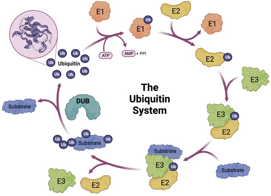 Figure 1. Ubiquitination cascade: E1 activation, E2 conjugation, E3 ligation, and proteasomal degradation. (Kennedy et al., 2022)
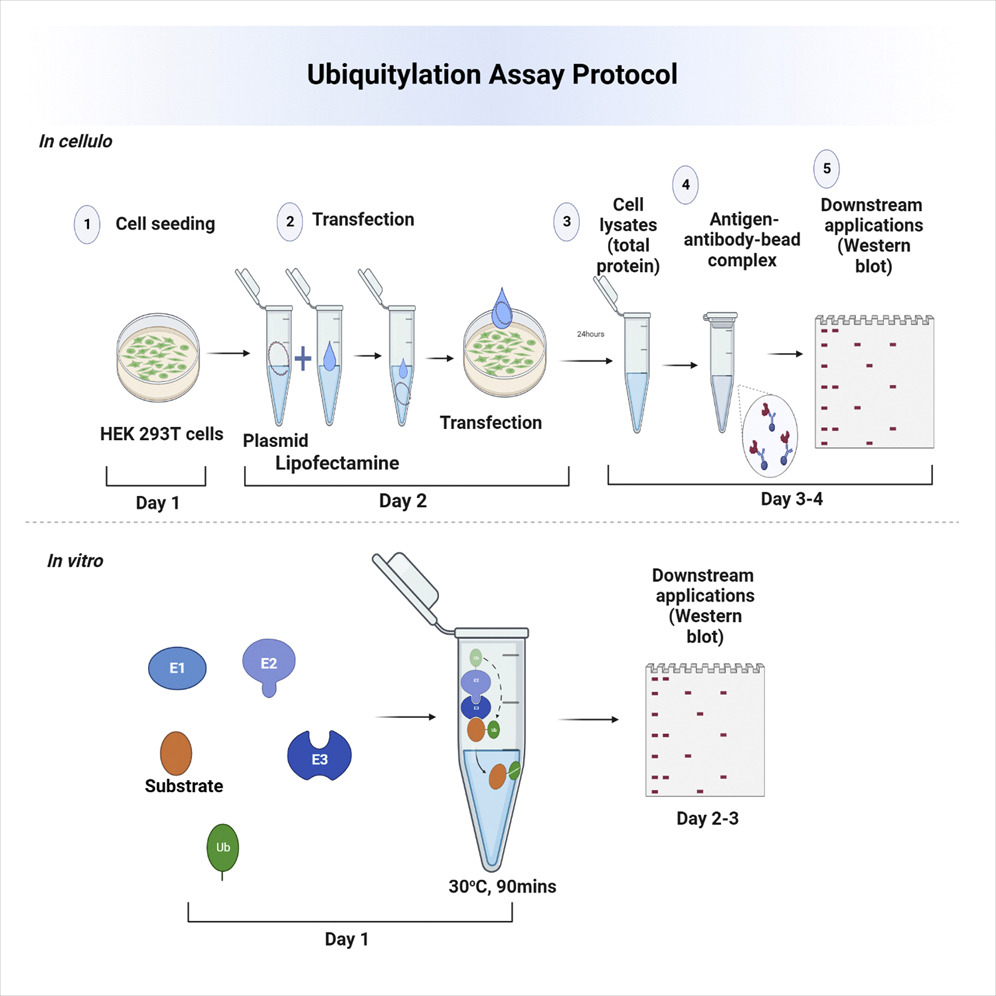 Figure 1. Protocol to detect in vitro and in cell ubiquitylation. (Hussain et al., 2022)