We use cookies to understand how you use our site and to improve the overall user experience. This includes personalizing content and advertising. Read our
Privacy Policy
Competitive binding assays represent a cornerstone of quantitative immunoassay methodology, enabling precise measurement of ligand-receptor and antibody-antigen interactions through the displacement of a labeled tracer by an unlabeled competitor. When implemented in the enzyme-linked immunosorbent assay (ELISA) format, competitive binding provides exceptional sensitivity, robustness, and scalability for applications ranging from therapeutic antibody characterization to biomarker quantification and immunogenicity assessment. Profacgen offers comprehensive Competitive Binding Assay (ELISA) services that leverage classical immunochemical principles alongside advanced detection platforms to deliver quantitative competition parameters, epitope mapping insights, and neutralizing antibody activity profiles. Our integrated approach supports drug development programs, diagnostic validation, and mechanistic immunology studies with rigorous statistical rigor and regulatory-aligned documentation.
Background: Principles of Competitive Binding ELISA
Competitive ELISA operates on the principle that an unlabeled analyte (competitor) and a labeled tracer compete for a limited number of binding sites on a capture molecule—typically an antibody, receptor extracellular domain, or ligand—immobilized on a solid-phase surface. As competitor concentration increases, tracer binding proportionally decreases, generating a sigmoidal dose-response curve from which quantitative binding parameters are extracted.
Figure 1. Typical sigmoidal calibration curve for a competitive ELISA using the 4-parameter logistic model. (Findlay and Dillard, 2007)
Binding Models and Quantitative Parameters
Profacgen employs mathematically rigorous models for competitive binding data analysis, ensuring accurate parameter extraction across diverse assay configurations:
Parameter
Definition
Calculation Method
Applications
IC50
Competitor concentration producing 50% inhibition of tracer binding under specific assay conditions
Four-parameter logistic regression of normalized signal versus log[competitor]; dependent on tracer concentration and affinity
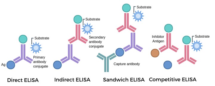
Competitive ELISA can be implemented in multiple orientations depending on the molecular system under investigation:
Direct Competitive ELISA: Competitor and enzyme-labeled antigen compete for binding to immobilized antibody. Applied when purified antigen is available for conjugation and antibody specificity is established.
Indirect Competitive ELISA: Competitor antigen displaces a primary antibody from immobilized antigen, with signal detected by enzyme-labeled secondary antibody. Eliminates need for antigen conjugation but requires higher antibody specificity.
Competitive Receptor Binding Assay: Soluble receptor extracellular domain immobilized on plate; biotinylated ligand tracer competed by unlabeled ligands, antibodies, or therapeutic candidates. Enables receptor pharmacology and antibody blocking assessment.
Sandwich Competitive ELISA: Capture antibody immobilized; competitor and labeled antigen compete for binding, with detection enhanced by a secondary detection antibody. Maximizes specificity for complex matrices such as serum or cell culture supernatant.
What We Offer
Profacgen provides a comprehensive portfolio of competitive binding ELISA services tailored to antibody therapeutics development, immunogenicity assessment, and molecular interaction characterization.
Antibody Competition and Epitope Mapping
Quantitative assessment of antibody-antigen binding site relationships through competitive displacement profiling:
Epitope Binning and Overlap Analysis
Pairwise competitive displacement of labeled reference antibodies by unlabeled test antibodies to classify epitope clusters (bins). Antibodies competing for the same epitope show complete displacement; non-competing antibodies bind distinct, non-overlapping sites. Results guide combination therapy design and intellectual property strategy.
Epitope Fine Mapping
Competition against overlapping peptide libraries, domain deletion mutants, or alanine-scanning variants to localize antibody binding to specific antigen regions. Combined with structural data, this enables rational antibody humanization and affinity maturation.
Cross-Reactivity Profiling
Competitive displacement across species orthologs, isoforms, and related family members to assess antibody selectivity. Critical for preclinical species selection, toxicology study design, and diagnostic specificity validation.
Affinity Maturation Monitoring
Comparative IC50 and Ki determination for antibody variants during directed evolution campaigns, providing quantitative feedback on selection pressure efficacy and guiding library design iterations.
Neutralizing Antibody Analysis
Functional assessment of antibody or serum-mediated inhibition of ligand-receptor interactions, enzymatic activity, or viral entry:
Anti-Drug Antibody (ADA) Neutralization: Competitive ELISA measuring patient serum ADA ability to block therapeutic antibody binding to its target antigen. Distinguishes binding ADAs from neutralizing ADAs with clinical relevance to therapeutic efficacy loss and safety signals.
Vaccine Efficacy Serology: Quantification of neutralizing antibody titers in immunized subjects by competition against labeled viral spike protein or receptor-binding domain tracers. Supports immunogenicity assessment and correlate-of-protection determination.
Receptor Blocking Assays: Evaluation of therapeutic antibody or antagonist ability to prevent ligand binding to cell surface or soluble receptors. Directly predictive of in vivo pharmacodynamic activity.
Enzyme Inhibition Profiling: Competitive displacement of substrate or cofactor binding to characterize mechanism-based inhibitors and allosteric modulators.
Detection Platform Options
Profacgen configures competitive ELISA detection to match sensitivity requirements, throughput constraints, and matrix compatibility:
Colorimetric ELISA: Horseradish peroxidase (HRP) or alkaline phosphatase (AP) conjugates with chromogenic substrates (TMB, pNPP). Standard sensitivity, broad dynamic range, cost-effective for high-throughput screening.
Chemiluminescent ELISA: Enhanced luminol or dioxetane substrates providing 10–100× sensitivity improvement over colorimetric formats. Ideal for low-abundance analytes, serum matrix interference, and regulatory compliance.
Fluorescent ELISA: Europium cryptate or dye-labeled detection reagents with time-resolved or conventional fluorescence detection. Supports multiplexing, low background signal, and homogeneous (no-wash) assay formats.
Alphascreen/AlphaLISA: Bead-based proximity luminescence enabling homogeneous competitive binding in solution without washing steps. Miniaturizable to 384- and 1536-well formats for ultra-high-throughput screening.
Service Procedure
Profacgen executes competitive binding ELISA projects through a standardized workflow ensuring assay robustness, quantitative accuracy, and regulatory compliance.
Assay Design and Tracer Selection: Evaluation of antigen-antibody system to determine optimal assay orientation (direct, indirect, sandwich); selection of high-affinity tracer with validated specificity; conjugation chemistry optimization (HRP, biotin, fluorophore, europium) preserving binding activity
Immobilization and Blocking Optimization: Capture molecule coating density titration; blocking buffer screening (BSA, casein, fish gelatin, proprietary formulations) to minimize nonspecific binding while preserving specific signal; plate type selection (high-binding polystyrene, streptavidin-coated, protein A/G-coated) matched to capture chemistry
Competitor and Tracer Concentration Optimization: Tracer KD determination by saturation binding; competitor IC50 pilot titration; selection of tracer concentration at 80% of saturation to maximize dynamic range; DMSO and vehicle tolerance assessment
Assay Validation and QC Implementation: Inter- and intra-assay precision (CV < 15%); Z'-factor determination for screening suitability (>0.5); reference compound IC50 confirmation against historical data; hook effect assessment for prozone detection in sandwich formats; parallelism testing for relative potency assays
Sample Analysis and Data Generation: Dose-response curves with 10–12 point titrations in duplicate or triplicate; matrix effect evaluation for serum, plasma, or cell culture supernatant; minimum required dilution (MRD) determination; four-parameter logistic or five-parameter logistic regression fitting
Quantitative Reporting and Interpretation: IC50, Ki, relative potency, and % neutralization reporting with 95% confidence intervals; epitope binning matrix visualization; structure-activity relationship correlation; regulatory-compliant study reports with full audit trails
Comprehensive Epitope Characterization: Integrated epitope binning, fine mapping, and cross-reactivity profiling within a single service provider, accelerating antibody engineering and intellectual property decision-making
Neutralizing Antibody Discrimination: Distinguishing binding versus neutralizing ADA with validated correlation to in vivo pharmacodynamic loss, supporting immunogenicity risk assessment and therapeutic monitoring strategies
Multi-Platform Detection Flexibility: Colorimetric, chemiluminescent, fluorescent, and bead-based homogeneous formats enabling sensitivity and throughput optimization for each project phase
Advanced Quantitative Modeling: Beyond IC50 reporting, we provide Ki determination, allosteric parameter extraction, and global fitting of multi-tracer datasets for mechanistic insight
Regulatory-Aligned Operations: GLP-compliant assay validation (ICH Q2(R1)), reference standard qualification, stability indication protocols, and complete documentation supporting IND, BLA, and diagnostic submission
Integrated Therapeutic Development Support: Seamless coordination with upstream antibody discovery, downstream cell-based functional assays, and pharmacokinetic/pharmacodynamic studies within Profacgen's service ecosystem
Representative Case Studies
Case 1: Epitope Binning and Combinatorial Pairing Strategy for Anti-SARS-CoV-2 Therapeutic Antibodies
Background:
A biotechnology company developed a panel of neutralizing antibodies against the SARS-CoV-2 receptor-binding domain (RBD) for emergency therapeutic deployment. With multiple candidates advancing in parallel, the team required rapid epitope classification to identify non-competing pairs suitable for cocktail therapy—an approach designed to prevent viral escape and extend therapeutic durability against emerging variants.
Our Solution:
Profacgen implemented a high-throughput competitive binding epitope binning platform using biotinylated RBD captured on streptavidin-coated plates. A reference antibody from each candidate was HRP-conjugated as tracer; all unlabeled candidates (n = 24) were tested as competitors at 10 µg/mL. Complete displacement (>80% tracer inhibition) indicated epitope overlap; partial or no displacement indicated distinct epitope bins. Selected non-competing pairs were further validated by surface plasmon resonance (SPR) to confirm simultaneous RBD binding.
Final Results:
The competitive ELISA binning resolved 24 antibodies into 6 distinct epitope clusters. Three clusters targeted the ACE2-binding site (competing with ACE2-Fc tracer), two targeted non-ACE2 overlapping cryptic epitopes, and one targeted a distal epitope accessible only in the RBD "up" conformation. A cocktail combining one antibody from the ACE2-competing cluster and one from the distal epitope cluster demonstrated synergistic neutralization against Alpha, Delta, and Omicron variants in pseudovirus assays. The epitope binning data supported the client's EUA submission and informed second-generation pan-variant antibody engineering.
Case 2: Anti-Drug Antibody Neutralization Assessment for a PEGylated Therapeutic Enzyme
Background:
A rare disease therapeutic program employed a PEGylated recombinant enzyme administered chronically. During Phase II, unexpected efficacy decline in 12% of patients correlated with rising anti-drug antibody (ADA) titers detected by a bridging ELISA. However, the bridging assay could not distinguish binding ADAs (non-neutralizing) from neutralizing ADAs functionally blocking enzyme activity—information critical for dose adjustment, retreatment decisions, and regulatory safety reporting.
Our Solution:
Profacgen developed a competitive neutralizing ADA assay: immobilized substrate was incubated with patient serum, followed by addition of limited enzyme tracer. Neutralizing ADAs in serum prevented enzyme-substrate binding, reducing signal proportionally to neutralizing titer. The assay was validated against a panel of characterized monoclonal antibodies with known neutralizing (enzyme-blocking) and non-neutralizing (PEG-specific) specificities. Results were reported as % neutralization and neutralizing titer (dilution achieving 50% signal inhibition).
Final Results:
Competitive neutralization analysis revealed that only 40% of ADA-positive patients (5% of total cohort) harbored neutralizing antibodies; the remaining 60% had non-neutralizing PEG-specific antibodies without functional enzyme inhibition. This stratification enabled targeted intervention: neutralizing ADA patients were switched to an alternative therapy, while non-neutralizing patients continued treatment with enhanced monitoring. The competitive assay was incorporated into the Phase III protocol as a biomarker-guided treatment algorithm, improving overall response rates and supporting regulatory approval with a differentiated risk-mitigation strategy.
Q: What is the difference between competitive ELISA and sandwich ELISA for antibody detection?
A: Competitive ELISA measures the ability of an unlabeled analyte to displace a labeled tracer from a capture molecule, generating an inverse signal relationship (more competitor = less signal). It is ideal for small molecules, haptens, and detecting antibodies that compete for a specific binding site. Sandwich ELISA uses two antibodies recognizing distinct epitopes to capture and detect the analyte, generating a direct signal relationship (more analyte = more signal). Competitive ELISA is preferred when: (1) only one specific antibody is available; (2) the analyte is too small for dual-antibody sandwich capture; (3) epitope overlap or blocking activity is the parameter of interest; or (4) detecting antibodies against a specific functional domain rather than total antibody titer.
Q: How does epitope binning by competitive ELISA differ from structural epitope mapping?
A: Epitope binning by competitive ELISA is a functional classification: antibodies that compete for the same tracer binding are assigned to the same bin, indicating spatial proximity or steric hindrance, but not necessarily identical amino acid contacts. Structural mapping (X-ray crystallography, cryo-EM, hydrogen-deuterium exchange MS) defines the precise residues involved in binding. Competitive binning is rapid, requires no purified antigen-antibody complex, and scales to large antibody panels. Structural mapping provides atomic-resolution detail but is low-throughput and resource-intensive. Profacgen recommends competitive binning for initial antibody clustering and structural mapping for lead candidate epitope definition.
Q: Can competitive ELISA distinguish neutralizing from non-neutralizing antibodies?
A: Yes, when the competitive tracer is a functional ligand or therapeutic antibody whose biological activity is known. If a test antibody displaces the tracer and the tracer's function is neutralized, the test antibody is classified as neutralizing. If the test antibody binds the antigen without displacing the functional tracer (detected by a separate binding assay), it is non-neutralizing. Profacgen designs competitive neutralization assays using enzyme substrates, receptor ligands, or viral entry probes as tracers to directly correlate displacement with functional blockade. This is critical for immunogenicity assessment, where only neutralizing anti-drug antibodies impact therapeutic efficacy.
Q: What factors affect IC50 reproducibility in competitive ELISA?
A: IC50 is an operational parameter dependent on assay conditions, not a true thermodynamic constant. Key variables include: (1) tracer concentration relative to its KD—higher tracer concentrations increase apparent IC50; (2) incubation time and temperature—insufficient equilibration leads to time-dependent IC50 shifts; (3) capture molecule density—overcrowding can cause avidity artifacts or steric hindrance; (4) matrix effects—serum proteins, lipids, and anti-coagulants can interfere with binding; (5) plate type and coating efficiency—batch variability affects signal range. Profacgen controls these variables through rigorous SOPs, reference compound tracking, and tracer KD confirmation in each assay run. For cross-study comparison, we recommend reporting Ki calculated by the Cheng-Prusoff equation rather than raw IC50.
Q: How is relative potency determined in competitive ELISA, and why is it important for biosimilars?
A: Relative potency compares the activity of a test sample to a reference standard under identical assay conditions, expressed as a percentage. It is determined by parallel-line analysis of log-dose response curves or by direct IC50 ratio comparison with confidence interval calculation. For biosimilar development, regulatory agencies require demonstration that the biosimilar is highly similar to the reference product with no clinically meaningful differences. Competitive binding relative potency provides quantitative evidence of functional comparability in the antigen-binding domain, complementing physicochemical and cell-based bioassay data. Profacgen's relative potency assays include parallelism testing, reference standard qualification, and statistical evaluation against predefined equivalence margins.
Q: What is the typical project timeline for competitive binding ELISA development?
A: Standard timelines are: (1) 4–6 weeks for direct competitive ELISA with available antibodies and antigens, including tracer conjugation, assay optimization, and pilot validation; (2) 6–8 weeks for complex systems requiring antibody generation, epitope validation, or matrix interference resolution; (3) 8–10 weeks for epitope binning panels with 20+ antibodies, including pairwise competition matrix generation and clustering analysis; and (4) 10–12 weeks for full GLP-compliant validation with documented precision, accuracy, specificity, robustness, and stability. High-throughput screening campaigns on validated assays require 2–3 weeks for single-point primary screens and 4–6 weeks for multi-point IC50 confirmation. Neutralizing ADA assay development from serum panel characterization to validated clinical testing typically requires 8–12 weeks.
References:
Findlay JWA, Dillard RF. Appropriate calibration curve fitting in ligand binding assays. AAPS J. 2007;9(2):E260-E267. doi:10.1208/aapsj0902029
Online Inquiry
Fill out this form and one of our experts will respond to you within one business day.
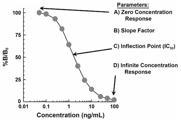 Figure 1. Typical sigmoidal calibration curve for a competitive ELISA using the 4-parameter logistic model. (Findlay and Dillard, 2007)