We use cookies to understand how you use our site and to improve the overall user experience. This includes personalizing content and advertising. Read our
Privacy Policy
Reporter gene assays represent a cornerstone of cell-based functional analysis, enabling quantitative readout of signal transduction pathway activation, transcription factor activity, and receptor-mediated gene regulation in living cells. By coupling a promoter element of interest to an easily detectable reporter enzyme—most commonly firefly or nanoluciferase luciferase—researchers can monitor cellular responses to ligands, therapeutic candidates, or genetic perturbations with exceptional sensitivity and dynamic range. These assays bridge molecular binding events to functional cellular outcomes, providing critical evidence of biological activity that complements biochemical interaction data. Profacgen offers comprehensive Reporter Gene Assay services leveraging optimized luciferase systems, validated pathway-specific reporter constructs, and high-throughput detection platforms to support drug discovery, therapeutic potency assessment, and mechanistic cell biology across NF-κB, STAT, Wnt/β-catenin, and other major signaling pathways.
Introduction: Assay Principle, Workflow, and Biological Meaning
Assay Principle
Reporter gene assays operate on the principle that transcriptional activation of a specific promoter or enhancer element can be quantified by coupling it to a reporter gene whose protein product generates a measurable signal. The typical configuration involves:
Promoter/Response Element: A cis-regulatory DNA sequence recognized by the transcription factor or pathway under investigation (e.g., NF-κB consensus binding sites, STAT-binding GAS elements, CREB response elements, TCF/LEF binding sites for Wnt signaling)
Reporter Gene: Firefly luciferase (Photinus pyralis), renilla luciferase (Renilla reniformis), or nanoluciferase (Oplophorus gracilirostris) cloned downstream of the response element, enabling luminescent signal generation proportional to transcriptional activity
Internal Control: A constitutively expressed reporter (typically renilla luciferase driven by SV40 or TK promoter) for normalization of transfection efficiency, cell number, and viability variations
Upon pathway activation, the transcription factor translocates to the nucleus, binds its cognate response element, and drives reporter gene transcription. The accumulated reporter protein catalyzes substrate oxidation, producing photons that are quantified by a luminometer. The normalized luciferase ratio (firefly/renilla) provides a robust, internally controlled measure of pathway-specific transcriptional activity.
Figure 1. A diagram of a how a reporter gene is used to study a regulatory sequence.
Workflow
Profacgen executes reporter gene assays through a standardized, quality-controlled workflow:
Cell Line Selection and Engineering: Selection of physiologically relevant cell backgrounds (HEK293, HepG2, U2OS, primary cells) or generation of stable reporter cell lines through lentiviral transduction of promoter-reporter constructs; monoclonal selection for uniform expression and response consistency
Reporter Construct Validation: Confirmation of construct integrity by sequencing; validation of basal reporter activity and dynamic range using known pathway activators and inhibitors; assessment of response linearity, fold-induction, and Z'-factor for screening suitability
Compound Treatment and Stimulation: Serial dilution of test compounds in appropriate vehicle; co-treatment with pathway-specific ligands (TNF-α for NF-κB, IFN-γ for STAT1, Wnt3a for β-catenin/TCF); incubation under optimized conditions (time, temperature, serum concentration) to achieve maximal signal-to-noise
Reporter Detection and Quantification: Reporter Detection and Quantification: Cell lysis followed by sequential luciferase reporter assay using firefly substrate (D-luciferin) and renilla substrate (coelenterazine); sequential luminescence measurement on plate-based luminometers; calculation of normalized firefly/renilla ratios and fold-induction over vehicle control.
Data Analysis and Interpretation: Dose-response curve fitting (four-parameter logistic) for EC50/IC50 determination; pathway specificity confirmation using orthogonal inhibitors; mechanism-of-action classification (agonist, antagonist, inverse agonist); correlation with orthogonal pathway readouts (Western blot for phospho-proteins, qPCR for endogenous target genes)
Biological Meaning
Reporter gene assays provide functional readouts that reflect the integrated activity of entire signaling cascades rather than isolated molecular interactions. This integration captures:
Pathway Fidelity: The complete sequence from receptor activation through second messenger generation, kinase cascade amplification, transcription factor modification, nuclear import, and gene transcription—revealing pathway integrity and cross-talk that biochemical assays miss
Cellular Context: Native expression of scaffolding proteins, negative regulators, and competing pathways that modulate signal amplitude and duration in physiologically relevant ways
Therapeutic Relevance: Functional consequences of drug binding (agonism, antagonism, inverse agonism, partial agonism) that predict in vivo pharmacological behavior more accurately than binding affinity alone
Temporal Dynamics: Time-dependent pathway activation and desensitization patterns that inform dosing regimens and sustained versus pulsatile therapeutic strategies
Applications
Reporter gene assays serve critical roles across therapeutic discovery, potency assessment, and mechanistic investigation:
GPCR Drug Discovery: Pathway-biased ligand identification by coupling Gs, Gi, or Gq-responsive CRE, SRE, or NFAT reporters to specific receptor subtypes; discrimination of balanced versus biased agonists
Cytokine and Growth Factor Signaling: Quantification of JAK-STAT pathway activation by interleukins, interferons, and growth factors; assessment of JAK inhibitor selectivity across STAT1, STAT3, and STAT5 reporters
Immunomodulatory Drug Development: NF-κB and AP-1 reporter assays for anti-inflammatory compound screening; evaluation of Toll-like receptor (TLR), NOD-like receptor (NLR), and RIG-I-like receptor (RLR) pathway modulators
Oncology Target Validation: Wnt/β-catenin, Hedgehog, and Notch pathway reporter assays for developmental pathway inhibitors; hypoxia-inducible factor (HIF) reporter assays for tumor microenvironment targeting
Biosimilar and Therapeutic Potency Testing: Quantitative comparison of biologic activity between innovator and biosimilar; batch release testing with reference standard calibration; stability indication under stress conditions
Toxicology and Safety Pharmacology: Off-target pathway activation screening (hERG, glucocorticoid receptor, androgen receptor reporters) to predict adverse effects early in development
Service Capabilities
Profacgen provides a comprehensive suite of reporter gene assay services with validated pathways, customizable constructs, and multi-format detection options.
Sequential firefly and renilla luciferase detection using automated plate luminometers; broad dynamic range (7–8 orders of magnitude), high sensitivity (attomolar detection), and robust signal normalization.
Bright Luciferase Reporter Systems
150-fold brighter than firefly luciferase with reduced ATP dependence; suitable for low-expression promoters, single-cell analysis, and miniaturized high-throughput formats.
Live-Cell Kinetic Reporters
Stable integration of luciferase reporters with real-time substrate delivery systems; continuous monitoring of pathway activation dynamics over hours to days
Multiplex Reporter Panels
Simultaneous assessment of multiple pathways in a single well using spectrally distinct luciferase reporters (firefly, renilla, ultra-bright luciferase system) or fluorescent reporters (GFP, RFP)
Deliverables
Each reporter gene assay project includes comprehensive analytical documentation and expert interpretation:
Normalized luciferase activity (firefly/renilla ratio) with fold-induction over vehicle control
Dose-response curves with EC50, IC50, Emax, and Hill slope parameters determined by four-parameter logistic regression
Pathway specificity confirmation through orthogonal inhibitor co-treatment and off-target pathway screening
Mechanism-of-action classification: full agonist, partial agonist, antagonist, inverse agonist, or allosteric modulator
Z'-factor and signal-to-background ratio for assay quality assessment
Time-course data for kinetic activation and desensitization profiling
Raw luminescence values and intermediate calculations upon request
Publication-ready figures and regulatory-compliant study reports with full experimental documentation
Extensive Validated Pathway Panel: Pre-constructed, sequence-verified reporter constructs for NF-κB, STAT1/3/5, Wnt/β-catenin, Hedgehog, CREB, NRF2/ARE, and custom pathways with demonstrated dynamic range and pharmacological responsiveness
Optimized Luciferase Systems: Firefly, renilla, and high-sensitivity luciferase reporters selected for maximal signal-to-noise, minimal autoluminescence, and appropriate spectral separation for multiplex applications
High-Throughput Screening Execution: Automated liquid handling, 384- and 1536-well format compatibility, and integrated data analysis pipelines enabling 10,000+ compound weekly throughput
Physiologically Relevant Cell Backgrounds: Engineered reporter lines in HEK293, HepG2, U2OS, and primary cell-compatible formats; genome-edited endogenous pathway component knockouts for specificity validation
Integrated Mechanistic Validation: Correlation with Western blot phospho-protein analysis, qPCR of endogenous target genes, and orthogonal functional assays (cAMP, calcium, IP1) to confirm pathway fidelity
Biosimilar and Regulatory Support: Reference standard qualification, relative potency determination, and stability indication protocols aligned with ICH guidelines and FDA biosimilar guidance
Representative Case Studies
Case 1: JAK Inhibitor Selectivity Profiling Using STAT Reporter Panel
Background:
A pharmaceutical company developing JAK inhibitors for autoimmune disease required comprehensive selectivity assessment across the four JAK isoforms (JAK1, JAK2, JAK3, TYK2) and their downstream STAT substrates. Biochemical kinase assays provided IC50 values against purified enzymes but could not predict cellular pathway selectivity due to differences in endogenous JAK expression levels, receptor coupling efficiency, and feedback regulation.
Our Solution:
Profacgen established a multiplex STAT reporter panel in a common HEK293 background: STAT1-GAS-firefly for JAK1/2/3/TYK2 (IFN-γ stimulated), STAT3-SIE-firefly for JAK1/2 (IL-6 stimulated), and STAT5-β-casein-firefly for JAK2 (IL-2 stimulated). Each reporter line was validated for JAK isoform dependence using siRNA knockdown and isoform-selective reference inhibitors (tofacitinib, ruxolitinib, oclacitinib). The test compound panel (n = 24) was profiled at 10-point concentrations across all three reporters with parallel phospho-STAT Western blot confirmation.
Final Results:
The reporter panel revealed that compound 17, nominally JAK2-selective by biochemical assay (50-fold vs. JAK1), showed only 3-fold cellular selectivity for STAT5 over STAT3 due to high endogenous JAK1 expression enabling compensatory signaling. In contrast, compound 9 exhibited 25-fold STAT5 selectivity despite modest biochemical JAK2 preference, attributed to poor membrane permeability limiting JAK1 access. These cellular selectivity insights redirected lead optimization toward compound 9's chemotype, and the STAT reporter panel was incorporated into the candidate's regulatory pharmacology package as a clinically relevant selectivity biomarker.
Case 2: NF-κB Reporter Assay for Anti-Inflammatory Biosimilar Potency Testing
Background:
A biosimilar developer required a cell-based potency assay for an anti-TNF-α monoclonal antibody referencing a marketed originator. While binding assays (SPR, ELISA) demonstrated comparable TNF-α affinity, regulatory agencies demanded functional evidence of equivalent neutralizing activity. The TNF-α-mediated NF-κB pathway is the primary mechanism of action for this therapeutic class, making an NF-κB reporter assay the most pharmacologically relevant potency test.
Our Solution:
Profacgen developed and validated a stable HEK293-NF-κB-luciferase reporter line responsive to TNF-α with an EC50 of 0.8 ng/mL and a dynamic range of 50-fold induction. The assay was optimized for biosimilar comparison: 96-well format, 4-hour TNF-α stimulation, 0.5 ng/mL TNF-α (submaximal for sensitivity), and 24-hour antibody pre-incubation. Biosimilar and innovator were tested at 8 concentrations in quadruplicate across three independent runs. Relative potency was calculated by parallel-line analysis of log-dose response curves against a qualified reference standard.
Final Results:
The NF-κB reporter assay demonstrated equivalent neutralizing potency between biosimilar and innovator (relative potency = 102%, 95% CI: 94–111%), with parallelism confirmed (p > 0.05 for non-parallelism test). The assay achieved a Z'-factor of 0.72 and inter-assay CV of 8.3%, meeting regulatory acceptance criteria. The validated assay was transferred to the client's QC laboratory with full documentation, and the NF-κB reporter potency data supported successful FDA biosimilar approval as part of the analytical similarity package.
Q: What is the difference between transient and stable reporter gene assays?
A: Transient reporter assays involve plasmid transfection 24–48 hours before assay execution, offering rapid setup and flexibility for construct testing but suffering from high well-to-well variability due to uneven transfection efficiency. Stable reporter assays use cell lines with chromosomally integrated reporter constructs, providing uniform expression, consistent response, and assay reproducibility suitable for screening campaigns and regulatory studies. Profacgen employs transient formats for exploratory construct validation and stable formats for high-throughput screening, potency testing, and longitudinal studies. Stable lines are generated by lentiviral transduction followed by antibiotic selection and monoclonal isolation to ensure homogeneous reporter expression.
Q: How do reporter gene assays differ from endogenous gene expression analysis (qPCR, RNA-seq)?
A: Reporter gene assays measure the activity of an exogenous, amplified promoter construct, providing a sensitive, rapid, and high-throughput readout of transcription factor activity with minimal background. They are ideal for compound screening, pathway mapping, and potency quantification. Endogenous gene expression analysis measures native transcript levels, capturing the full complexity of chromatin regulation, enhancer involvement, and mRNA stability but with lower throughput and greater experimental complexity. Reporter assays may not fully recapitulate endogenous gene regulation due to missing distal enhancers, chromatin context, and feedback loops. Profacgen recommends reporter assays for primary screening and potency testing, with orthogonal qPCR validation of key endogenous target genes to confirm physiological relevance.
Q: Can reporter assays distinguish between direct pathway activation and indirect cellular stress responses?
A: Yes, through rigorous specificity controls. Non-specific cellular stress (cytotoxicity, oxidative stress, unfolded protein response) can artifactually activate certain reporters, particularly NF-κB and AP-1. Profacgen implements multiple discrimination strategies: (1) cell viability assessment parallel to reporter readout to exclude cytotoxic artifacts; (2) orthogonal pathway inhibitors to confirm mechanism-specificity (e.g., IKK inhibitors for NF-κB, JAK inhibitors for STAT); (3) dominant-negative pathway component co-expression to validate reporter dependence; (4) time-course analysis to distinguish rapid, specific activation from delayed stress responses; and (5) correlation with phospho-protein Western blots for direct pathway component modification. Compounds showing reporter activation without corresponding phospho-protein changes or with concurrent cytotoxicity are flagged as potential false positives.
Q: What are the advantages of your high-sensitivity luciferase system over conventional firefly luciferase in reporter assays?
A: High-sensitivity luciferase systems offer several technical advantages: (1) superior brightness (~150-fold vs. firefly luciferase), enabling detection of weak promoters and single-cell resolution; (2) smaller size (19 kDa vs. 61 kDa), reducing steric interference with fusion protein function; (3) ATP-independent catalysis, minimizing ATP concentration artifacts in metabolically stressed cells; (4) improved stability and reduced background luminescence; (5) compatibility with extracellular inhibitors for signal tuning and temporal control; and (6) spectral compatibility with fluorescent reporters for multiplex applications. The trade-off is higher reagent cost and the need for specialized luminometers with enhanced sensitivity. Profacgen selects luciferase systems based on promoter strength, dynamic range requirements, and multiplex configuration.
Q: How are reporter gene assays used for GPCR drug discovery?
A: GPCR signaling is routinely interrogated using pathway-specific reporters coupled to distinct G protein classes: CRE-luciferase for Gs-mediated cAMP elevation (β-adrenergic, glucagon, PTH receptors); SRE-luciferase for Gq-mediated calcium/PKC activation (α1-adrenergic, muscarinic M1/M3, histamine H1 receptors); and NFAT-luciferase or SRF-RE-luciferase for G12/13 and Gi/o pathways. Profacgen generates stable GPCR-reporter cell lines with receptor overexpression and pathway-specific reporters, enabling: (1) agonist EC50 determination and efficacy ranking; (2) antagonist IC50 and mode-of-action classification; (3) allosteric modulator characterization through probe-dependent shifts; and (4) biased agonism assessment by comparing potency ratios across multiple pathway reporters for the same receptor. These assays bridge biochemical binding data to functional pharmacology essential for therapeutic candidate selection.
Q: What is the typical project timeline for reporter gene assay development?
A: Standard timelines are: (1) 3–4 weeks for transient reporter assay execution using available validated constructs and cell lines; (2) 6–8 weeks for stable reporter cell line generation including lentiviral transduction, antibiotic selection, monoclonal isolation, and response validation; (3) 4–6 weeks for assay optimization including stimulation time, ligand concentration, cell density, and detection parameter determination; (4) 8–10 weeks for full assay validation with Z'-factor confirmation, reference compound qualification, and inter-assay precision assessment; and (5) 10–12 weeks for GLP-compliant validation with documented robustness, specificity, and stability testing. High-throughput screening campaigns on validated assays require 2–3 weeks for single-concentration primary screens and 4–6 weeks for multi-point dose-response confirmation. Custom pathway reporter construction from promoter design through validation typically requires 10–14 weeks.
References:
De Wet JR, Wood KV, DeLuca M, et al. Firefly luciferase gene: structure and expression in mammalian cells. Molecular and Cellular Biology. 1987;7(2):725-737.
Hall MP, Unch J, Binkowski BF, et al. Engineered luciferase reporter from a deep sea shrimp utilizing a novel imidazopyrazinone substrate. ACS Chemical Biology. 2012;7(11):1848-1857.
Online Inquiry
Fill out this form and one of our experts will respond to you within one business day.
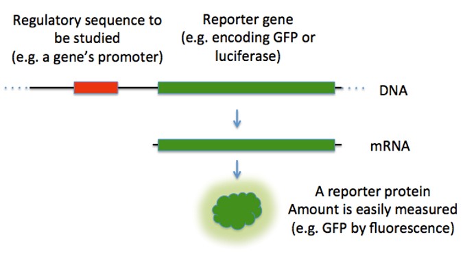 Figure 1. A diagram of a how a reporter gene is used to study a regulatory sequence.